Repro.ucsd.edu

Androgen responsiveness of the pituitary gonadotrope cell line L T2
M A Lawson, D Li, C A Glidewell-Kenney and F J López
Department of Pharmacology, Ligand Pharmaceuticals Inc., 10275 Science Center Drive, San Diego, California 92121, USA
(Requests for offprints should be addressed to F J López) (M A Lawson is now at Department of Reproductive Medicine, School of Medicine, University of California, San Diego, 9500 Gilman Drive, La Jolla, (C A Glidewell-Kenney is now at CLONTECH Laboratories, Inc., 1020 East Meadow Circle, Palo Alto, California 94303, USA) Abstract
Androgens have a profound effect on the hypothalamic– (MMTV) promoter, showed that a functional AR is also pituitary axis by reducing the synthesis and release of the pituitary gonadotropin LH. The effect on LH is partly a (DHT), 7 -methyl-19-nortestosterone (MENT), and consequence of a direct, steroid-dependent action on fluoxymesterone (FLUOXY) increased reporter gene pituitary function. Although androgen action has been activity in the rank order of potencies MENT>DHT> well studied in vivo, in vitro cell models of androgen action TEST>FLUOXY. Additionally, activation of MMTV on pituitary gonadotropes have been scarce. Recently, an promoter activity by DHT in L T2 cells was diminished LH-expressing cell line, L T2, was generated by tumori- by the AR antagonists casodex and 2-hydroxy-flutamide, genesis targeted to the LH-producing cells of the mouse indicating that the effects of DHT are mediated through pituitary. The purpose of these studies was to determine AR. In summary, these studies showed that the L T2 the presence of androgen receptor (AR) and establish its cell line is a useful model for the evaluation and function in this cell line. RT-PCR analysis indicated that molecular characterization of androgen action in pituitary the L T2 cell line expresses AR mRNA. Transient transfection assays, using the mouse mammary tumor virus Journal of Endocrinology (2001) 170, 601–607
Introduction
hormone, the secretagogue of the gonadotropins, as well asto estrogen (Turgeon et al. 1996, Schreihofer et al. 2000).
Androgen action is mediated by the androgen receptor In these studies, we have investigated the potential use of (AR) which modulates activation of specific genes by this cell line as a model to evaluate androgen action at the homodimeric AR bound to DNA, or by interactions with molecular level in pituitary gonadotropes. We have other transcription factors. Both positive and negative identified the presence of endogenous AR in the L T2 regulation of target genes is a characteristic mode of action cell line. We have also defined the response to various for steroid receptors (Beato et al. 1995). Androgens are androgens and assessed receptor specificity using AR clearly involved in reproductive tissue development, antagonists. These studies have demonstrated the presence muscle and bone homeostasis, sex behavior, metabolism, of a functional AR in the L T2 cell line and establish these etc. (see Mooradian et al. 1987 for a review). In the cells as a model for the evaluation of the molecular hypothalamic–pituitary axis, androgens negatively regulate mechanisms of androgen action in pituitary gonadotropes.
gonadotropin secretion (Kalra & Kalra 1983, Gharib et al.
1990) and, consequently, reproductive function.
Materials and Methods
Existing models for the in vitro analysis of steroid action on reproductive endocrine tissues are either laborious or of limited availability. Recently, a number of pituitary celllines have been developed using targeted tumorigenesis of L T2 cells were licensed from the University of pituitary gonadotropes in transgenic mice (Alarid et al. California, San Diego (La Jolla, CA, USA). Cells were 1996, 1998). One of these cell lines, L T2, expresses cultured in high glucose, HEPES-buffered Dulbecco’s luteinizing hormone (LH) (Turgeon et al. 1996) and follicle-stimulating hormone (FSH) (Graham et al. 1999) Walkersville, MD, USA) supplemented with 2 mM under specific culture conditions. It has been reported -glutamine and 10% fetal bovine serum (Hyclone, Logan, that this cell line is responsive to gonadotropin-releasing UT, USA), penicillin and streptomycin (Gibco BRL, Journal of Endocrinology (2001) 170, 601–607
Online version via http://www.endocrinology.org 0022–0795/01/0170–601  2001 Society for Endocrinology Printed in Great Britain 602 M A LAWSON and others · Androgen responsiveness of L T2 cells Grand Island, NY, USA). Cells were grown at 37 C in a humidified incubator in an atmosphere of 5% CO :95% L T2 cells were plated on Falcon primaria plates (Becton air. Subculture of L T2 cells was accomplished by gentle Dickinson, Franklin Lakes, NJ, USA) at a density of trypsinization when they approached 80% confluence.
50 000 cells/cm2 in phenol red-free, high glucose DMEM Subcultivation ratios of 1:5 to 1:20 were routinely used.
supplemented with 2 mM -glutamine and 10% charcoal/dextran-stripped fetal bovine serum (Hyclone), penicillin and streptomycin (GIBCO/BRL). After growth over-night, the cell medium was replaced with fresh medium L T2 cells were cultured to confluency and, after washing containing either vehicle or test compounds. Cells the monolayer with phosphate-buffered saline, total RNA were immediately transfected with the reporter plasmid was extracted by lysis in TRIzol reagent according to the pGL3-MMTV (mouse mammary tumor virus; 10 µg/ml), manufacturer’s instructions. Total RNA from mouse consisting of the firefly luciferase reporter gene from the prostate was also isolated and served as a positive control pGL3-basic reporter plasmid (Promega, Madison, WI, for AR mRNA. After alcohol precipitation, total RNA USA) under the transcriptional control of the androgen- pellets were dissolved in water and absorbance was sensitive MMTV LTR promoter element. As an internal measured at 260 and 280 nm. One microgram of total control, cells were co-transfected with the pCMV- - RNA from L T2 cells and mouse prostate was DNase galactosidase reporter plasmid (1 µg/ml) (CLONTECH treated and reverse transcribed using the superscript II kit Laboratories, Inc., Palo Alto, CA, USA). The FuGENE 6 (Gibco BRL) and priming with oligo-dT, following the manufacturer’s recommendations. cDNAs (equivalent to Indianapolis, IN, USA) was used for the transfection 1 µg reverse transcribed total RNA) were PCR amplified experiments following the manufacturer’s recommen- using a hot-start PCR protocol and AmpliTaq Gold dations. Cells were treated with vehicle or the various (PE Applied Biosystems, Foster City, CA, USA). The reactions were carried out in 100 µl volume of 1 (5 µl/well) and were incubated for 24 h. Thereafter, cells AmpliGold buffer II containing 1 mM MgCl were lysed with phosphate-buffered saline containing 400 nM of each primer. Samples were heated at 94 C for 14 min followed by successive heating steps at 94 C for activity were measured in the same aliquot using the dual 30 s, 52·5 C for 30 s and 72 C for 45 s. Thirty-five cycles light assay system (Tropix, Bedford, MA, USA) in a were used for amplification followed by a final extension LB96V luminometer (EG&G Berthold, Bad Wildbad, period at 72 C for 10 min. Primers for mouse AR Germany) controlled by the WinGlow software (EG&G (accession number M37890) (Gaspar et al. 1990, 1991, He Berthold). Luciferase reporter gene activity normalized to et al. 1990, Faber et al. 1991) used in the RT-PCR paradigm were as follows: forward primer: 5 -AACAACA GCAGCAGCACC-3 (bases 530 to 547), reverse primer5 -GGATTGGAAGGTGAGAGAGCTT-3 (bases 953to 934).
samples per group. The results were analyzed forstatistically significant differences by a one-way analysis Amplification products (1/10th of the PCR reaction) of variance and Dunnett’s post-hoc test on data either were resolved by electrophoresis through a 1% agarose gel untransformed or optimally transformed by the method of and transferred to a nylon membrane (Hybond-N+; Box–Cox (Box & Cox 1964) using the JMP statistical Amersham, Arlington Heights, IL, USA) using a trans-Vac analysis software (SAS Institute, Cary, NC, USA). Specific data transformations are mentioned in the figure legends.
Inc., San Francisco, CA, USA). The identity of the amplification products was confirmed by hybridization the four-parameter logistic equation on either untrans- using an oligonucleotide probe specific to the mouse AR formed or Box–Cox-transformed data by a previously described method (Ghosh et al. 1998). A P<0·05 was AAACA-3 ). The oligonucleotide was labeled with 32P designated as the minimum criterion for declaring using terminal transferase and the resulting probe was statistically significant differences.
diluted in Rapid-Hyb solution (Amersham). The blot washybridized to the diluted probe for 2 h at 42 C andwashed once at room temperature with 50 ml 5 0·1% SDS. Two additional stringency washes were per-formed at 42 C utilizing 1 To determine whether the pituitary cell line L T2 expresses the AR gene, RT-PCR was conducted on total Journal of Endocrinology (2001) 170, 601–607
Androgen responsiveness of L T2 cells · M A LAWSON and others 603 Although direct activation of AR leads to transcriptional responses dependent on the interaction of ligand-bound AR with a competent promoter element, othermechanisms for AR-dependent activation of transcriptionhave been reported that are resistant to inhibition by theandrogen receptor antagonists 2-hydroxy-flutamide andcasodex (Peterziel et al. 1999). Therefore, to establish thatthe androgen responsiveness of pGL3-MMTV is depen-dent on the transcription activation properties of ligand-bound AR, we tested the ability of the AR antagonists2-hydroxy-flutamide and casodex to block the DHT- 
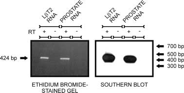
Figure 1 L T2 cells express AR mRNA. Primers specific for mouse
evoked response of the reporter gene in L T2 cells. Cells AR were used to amplify reverse transcribed mouse AR cDNA were treated with vehicle, 2 nM DHT (EC ) or 2 nM from L T2 cells or mouse prostate total RNA. Amplification DHT and increasing concentrations of 2-hydroxy- products were resolved by electrophoresis through a 1% agarosegel and Southern blotted with an oligonucleotide probe flutamide or casodex concurrently with transfection. Both corresponding to a sequence internal to the amplification primer 2-hydroxy-flutamide and casodex blocked DHT-induced symbols indicate the inclusion or omission of response of the reporter gene, indicating AR-dependent RT in the reverse transcription step prior to amplification. Size activity. In addition, both compounds demonstrated full indication is derived from a 100 bp ladder included in the agarose fficacy, since they suppressed DHT-mediated reporter gene response to levels equivalent to the untreated vehiclecontrols (Fig. 3). Although both AR antagonists were fullyefficacious, they exhibited a marked difference in potency RNA isolated from L T2 cells. Amplification products of (56 nM for 2-hydroxy-flutamide versus 1·2 µM for predicted mobility were identified in RT-PCR reactions that included reverse transcriptase in L T2 and prostatetotal RNA samples (Fig. 1; left panel). Reactions notcontaining reverse transcriptase did not yield any amplifi- Discussion
cation product (Fig. 1; left panel). These data indicate thatthe source for the target sequence is mRNA rather than The results of these studies indicate that the gonadotrope contaminating genomic DNA. As an additional verifi- cell line L T2 represents a valuable model for the analysis cation of RT-PCR fidelity, PCR products were blotted of androgen action in the pituitary that would facilitate the and probed with a radiolabeled oligonucleotide derived dissection of the molecular mechanisms involved in the from AR sequence internal to the primer sequences used regulation of gonadotropin production and secretion. Our in the PCR reaction. A correctly migrating band contain- data demonstrate that L T2 cells express AR mRNA and ing appropriate AR-derived sequences was observed (Fig.
functional protein as determined by activation of the 1; right panel). Thus, the L T2 cell line clearly expresses androgen-sensitive MMTV promoter in a transient trans- fection paradigm. DHT, FLUOXY, MENT and TEST The presence of functional AR in L T2 cells was elicit robust, two- to fourfold increases in reporter gene evaluated in transient transfection assays using an activity with the following rank of potencies: MENT> androgen-sensitive reporter. The pGL3-MMTV reporter DHT>TEST>FLUOXY. This response is AR depen- plasmid used in these studies containing the MMTV dent as evidenced by the blockade of DHT-evoked long terminal repeat (LTR) promoter driving luciferase activity of the reporter gene by the well-characterized AR gene expression was chosen because of the well- antagonists 2-hydroxy-flutamide and casodex. Our obser- documented sensitivity of the promoter to direct vations reinforce the notion that the gonadotrope itself AR action at the level of transcription (Beato et al. represents a potential site for androgen action in regulating gonadotropin synthesis and secretion.
(FLUOXY), 7 -methyl-19-nortestosterone (MENT), or Androgens are the predominant peripheral signal regu- testosterone (TEST) caused a dose-dependent increase in lating LH secretion by acting at both the hypothalamus pGL3-MMTV reporter gene activity in the L T2 cells (Damassa et al. 1976, Clayton et al. 1982) and pituitary (Fig. 2). The rank potencies for the compounds tested (Kingsley & Bogdanove 1973, Drouin & Labrie 1976, were MENT>DHT>TEST> FLUOXY and are sum- Giguere et al. 1981, Kotsuji et al. 1988) sites, although marized in Table 1. Induction of reporter activity at their mechanism of action in either tissue is not well maximal doses ranged approximately two- to fourfold.
defined. Some evidence has been presented to suggest that The dose-dependent increase in pGL3-MMTV reporter the hypothalamus alone is the site of androgen feedback gene expression in androgen-treated cells indicated that (Dubey et al. 1987, Tilbrook et al. 1991). With respect to the pituitary site of action, AR is expressed in the anterior Journal of Endocrinology (2001) 170, 601–607
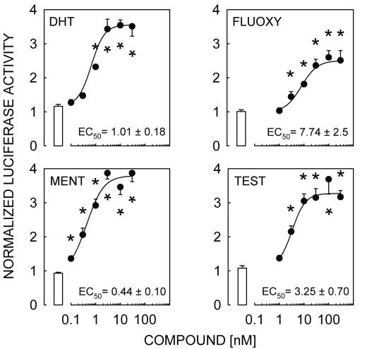
604 M A LAWSON and others · Androgen responsiveness of L T2 cells Figure 2 DHT, FLUOXY, MENT and TEST activate the pGL3-MMTV reporter transfected
into L T2 cells. Luciferase values are reported relative to -galactosidase activity as an
internal control. The open bar represents the vehicle-treated group for each treatment.
Results from one of three independent experiments are presented. Data for each group
correspond to the means
S.E.M. of six wells. Asterisks denote statistically significant differences versus the respective vehicle-treated groups. For statistical analysis, data wereconverted to the natural logarithm.
pituitary (Naess et al. 1975, Sar & Stumpf 1977, Thieulant 1999, Pelletier et al. 2000). In particular, AR-like & Pelletier 1979, Schanbacher et al. 1984, Bonsall et al. immunoreactivity has been identified in secretory cells of 1985, Sar et al. 1990, Kimura et al. 1993, Abdelgadir et al. the anterior pituitary (Sar et al. 1990, Pelletier et al. 2000) Table 1 Summary of potencies for androgens and antiandrogens when tested in multiple
experiments
95% Confidence
Interassay
estimate
coefficients of
variation (%)
Compound
Dihydrotestosterone
Data denote the results of (n) independent experiments using six replicates per group. The means arecalculated as the average potency estimate for each experiment weighted by the variance of eachexperiment. The first four compounds were tested alone, the last two were tested in the presence of anEC75 of DHT (2 nM). Weighted mean potency is reported Journal of Endocrinology (2001) 170, 601–607
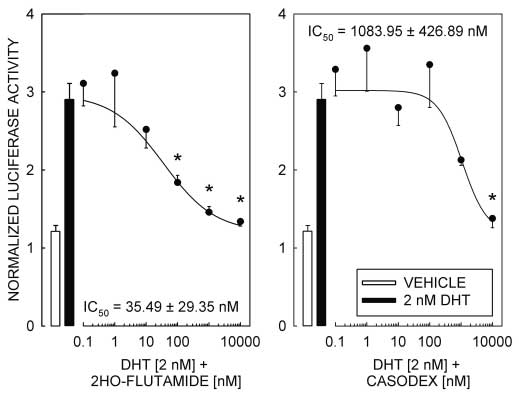
Androgen responsiveness of L T2 cells · M A LAWSON and others 605 Figure 3 AR antagonists 2-hydroxy-flutamide and casodex block DHT-induced reporter
gene expression in L T2 cells. Cells were co-transfected with the pGL3-MMTV and
pCMV- -galactosidase reporter genes. Transfected cells were concurrently treated with
vehicle (open bars), an EC75 dose of DHT (2 nM; solid bars) or DHT plus increasing
concentrations of 2-hydroxy-flutamide (2HO-FLUTAMIDE) or casodex (symbols andcurves). Results from one of two independent experiments are presented. Data for eachgroup are represented as the mean S.E.M. of six wells. Asterisks denote statistically significant differences versus the DHT-treated groups. For statistical evaluation, data wereelevated to and AR-positive staining has been described in human respectively (Drouin & Labrie 1976). In L T2 cells, DHT pituitary FSH- and LH-producing cells (Kimura et al. was also approximately 2·5 times more potent than TEST 1993). Similarly, morphological/biochemical studies have in inducing activation of the reporter gene, indicating that shown abundant androgen-binding sites in rat pituitary in these immortalized cells both steroids behave as gonadotropes (Sar & Stumpf 1977). Our data demonstrate observed in primary cultures of pituitary cells. In addition, the presence of AR mRNA and protein in immortalized it has been shown that MENT is 12–25 times more potent LH-secreting cells. The L T2 cell line therefore appears to than TEST in reducing orchidectomy-induced elevation have retained the ability to express AR as observed in the of LH secretion (Kumar et al. 1992). This difference in pituitary in vivo. The presence of functional AR in an potency is also observed in immortalized gonadotropes, immortalized, yet highly differentiated, cell line such as since MENT has an approximately tenfold lower ED50 L T2 cells provides a model system for addressing issues of than TEST in L T2 cells. Overall, these observations direct androgen action at the level of the pituitary. This support the notion that the L T2 cell line represents a model offers a simpler system where the confounding relevant model to assess androgen action in pituitary influence of other endocrine effectors normally found in vivo or in primary cultures of gonadotropes is absent.
The establishment of an in vitro cell model of gonado- Functional studies suggest profound effects of androgens trope function such as the L T2 cell line that is androgen in the modulation of LH secretion in primary cultures of responsive represents an important addition to the models pituitary cells. Both TEST and DHT have been shown to used to evaluate tissue-selective actions of androgens.
readily suppress LH-releasing hormone (LHRH)-induced Synthetic androgens and antiandrogens exhibit tissue sel- LH release from anterior pituitary cells in either static ectivity that results in different physiological consequences or dynamic culture conditions (Drouin & Labrie 1976, when administered in vivo. For example, both 2-hydroxy- Kotsuji et al. 1988). DHT is approximately three times flutamide and casodex cause the regression of seminal more potent than TEST in inhibiting LHRH-induced LH vesicles and ventral prostate in intact mature rats treated for 14 days; however, casodex does not cause significant Journal of Endocrinology (2001) 170, 601–607
606 M A LAWSON and others · Androgen responsiveness of L T2 cells changes in circulating levels of LH or TEST, whereas transcription factors binding to their own regulatory se- 2-hydroxy-flutamide does (Furr et al. 1987). In a pituitary quences in the promoters. Further examination of the cell background, casodex appears 20-fold less potent than flutamide in blocking DHT-induced MMTV promoter promoters is necessary to determine the mechanism by activity in L T2 cells. However, when tested in a which androgens inhibit transcriptional activity.
co-transfection assay in CV1 cells, Hamann et al. (1998) In summary, we have demonstrated that the L T2 cell line is a useful model for the study of androgen action in casodex respectively. This difference in activity may be the anterior pituitary. The availability of a highly differ- dependent on the cell background, suggesting that casodex entiated cell line with a consistent gonadotrope phenotype may be less effective in pituitary gonadotropes than facilitates the study of the mechanisms of androgen action flutamide in blocking androgen action. Our observations in the gonadotrope. The L T2 cells provide a consistent suggest that the interaction of AR with intracellular source of material for detailed examination of the tran- molecular targets may differ between the two antiandro- scriptional mechanisms of androgen action. Further study gens tested, leading to a decreased potency of the com- of AR action on endogenously expressed genes, as well as pound. Precedence for such an effect is found in the study of antiandrogen action via ARE-dependent and analysis of co-factor interaction of estrogen receptor with non-ARE, protein–protein interaction-dependent mech- estrogen receptor modulators (Wijayaratne et al. 1999).
anisms will lead to a better mechanistic understanding of In the case of the estrogen receptor, altered activity of AR as a modulator of reproductive functions.
the ligand-bound estrogen receptor can be correlatedto altered conformation of the receptor. Furthermore, Acknowledgements
degradation of AR, thereby exerting antiandrogen activity A portion of this work was supported by NIH grants by reducing receptor content (Veldscholte et al. 1992, R01HD-37568 and U54HD-12303 to M A L. We would Kemppainen & Wilson 1996, Waller et al. 2000). More like to thank Drs William Chang, Patricia Finn, William studies evaluating non-androgen response element (ARE)- Schrader, Tom Spady and Humberto Viveros for review dependent AR activity on the LH promoter are required Androgen-dependent reduction of LH secretion via direct action at the pituitary, rather than at the hypothal- References
amus, could be explained by modulation of LHRH-receptor function, LH production, or both. Evidence for Abdelgadir SE, Roselli CE, Choate JV & Resko JA 1999 Androgen receptor messenger ribonucleic acid in brains and pituitaries of male an androgen-dependent reduction in pituitary LHRH rhesus monkeys: studies on distribution, hormonal control, and receptors has been demonstrated by Giguere et al. (1981).
relationship to luteinizing hormone secretion. Biology of Reproduction In agreement with this, pituitary cells from castrated rats 60 1251–1256.
show increased responsiveness to an LHRH challenge in Alarid ET, Windle JJ, Whyte DB & Mellon PL 1996 Immortalization vitro (O’Conner et al. 1980). Since, in functional studies of pituitary cells at discrete stages of development by directed
oncogenesis in transgenic mice. Development 122 3319–3329.
using primary pituitary cell cultures, androgens do not Alarid ET, Holley S, Hayakawa M & Mellon PL 1998 Discrete stages alter basal LH secretion (Drouin & Labrie 1976, Kotsuji of anterior pituitary differentiation recapitulated in immortalized cell et al. 1988), it has been suggested that an important lines. Molecular and Cellular Endocrinology 140 25–30.
mechanism of action for androgens in the control of LH Beato M, Herrlich P & Schutz G 1995 Steroid hormone receptors: many actors in search of a plot. Cell 83 851–857.
secretion in gonadotropes is the reduction of LHRH Beato M, Truss M & Chavez S 1996 Control of transcription by receptors rather than direct modulation of LH production.
steroid hormones. Annals of the New York Academy of Sciences 784
More recent studies have suggested that AR can suppress Bonsall RW, Rees RH & Michael RP 1985 The distribution, nuclear ligand-dependent fashion using multiple mechanisms.
uptake and metabolism of [3H]dihydrotestosterone in the brain,pituitary gland and genital tract of the male rhesus monkey. Journal of Steroid Biochemistry 23 389–398.
been described (Clay et al. 1993). Mutational analysis of Box GEP & Cox DR 1964 An analysis of transformations. Journal of the ARE has indicated that this sequence is not required the Royal Statistical Society Series B 26 211–252.
for liganded AR-dependent repression of the promoter Clay CM, Keri RA, Finicle AB, Heckert LL, Hamernik DL, Marschke KM, Wilson EM, French FS & Nilson JH 1993 (Heckert et al. 1997). In addition, two elements, the Transcriptional repression of the glycoprotein hormone alpha basal- and tandem cAMP-regulatory elements, have been subunit gene by androgen may involve direct binding of androgen receptor to the proximal promoter. Journal of Biological Chemistry AR-mediated suppression of transcription (Heckert et al. 268 13556–13564.
1997). Therefore, the AR-dependent inhibitory effect of Clayton RN, Popkin RM & Fraser HM 1982 Hypothalamic regulation of pituitary gonadotropin-releasing hormone receptors: effects of gonadotropin-releasing hormone immunoneutralization.
protein–protein interactions between AR and other Endocrinology 110 1116–1122.
Journal of Endocrinology (2001) 170, 601–607
Androgen responsiveness of L T2 cells · M A LAWSON and others 607 Damassa DA, Kobashigawa D, Smith ER & Davidson JM 1976 Kingsley TR & Bogdanove EM 1973 Direct feedback of androgens: Negative feedback control of LH by testosterone: a quantitative localized effects of intrapituitary implants of androgens on study in male rats. Endocrinology 99 736–742.
gonadotrophic cells and hormone stores. Endocrinology 93
Drouin J & Labrie F 1976 Selective effect of androgens on LH and FSH release in anterior pituitary cells in culture. Endocrinology 98
Kotsuji F, Winters SJ, Attardi B, Keeping HS, Oshima H & Troen P 1988 Effects of gonadal steroids on gonadotropin secretion in males: Dubey AK, Zeleznik AJ & Plant TM 1987 In the rhesus monkey studies with perifused rat pituitary cells. Endocrinology 123
(Macaca mulatta), the negative feedback regulation of follicle- stimulating hormone secretion by an action of testicular hormone Kumar N, Didolkar AK, Monder C, Bardin CW & Sundaram K 1992 directly at the level of the anterior pituitary gland cannot be The biological activity of 7 alpha-methyl-19-nortestosterone is not accounted for by either testosterone or estradiol. Endocrinology 121
amplified in male reproductive tract as is that of testosterone.
Endocrinology 130 3677–3683.
Faber PW, King A, van Rooij HC, Brinkmann AO, de Both NJ & Mooradian AD, Morley JE & Korenman SG 1987 Biological actions of Trapman J 1991 The mouse androgen receptor. Functional analysis androgens. Endocrine Reviews 8 1–28.
of the protein and characterization of the gene. Biochemical Journal Naess O, Hansson V, Djoeseland O & Attramadal A 1975 278 269–278.
Characterization of the androgen receptor in the anterior pituitary
of the rat. Endocrinology 97 1355–1363.
Furr BJ, Valcaccia B, Curry B, Woodburn JR, Chesterson G & O’Conner JL, Allen MB & Mahesh VB 1980 Castration effects on the Tucker H 1987 ICI 176,334: a novel non-steroidal, peripherally response of rat pituitary cells to luteinizing hormone-releasing selective antiandrogen. Journal of Endocrinology 113 R7–R9.
hormone: retention in dispersed cell culture. Endocrinology 106
Gaspar ML, Meo T & Tosi M 1990 Structure and size distribution of the androgen receptor mRNA in wild-type and Tfm/Y mutant Pelletier G, Labrie C & Labrie F 2000 Localization of oestrogen mice. Molecular Endocrinology 4 1600–1610.
receptor alpha, oestrogen receptor beta and androgen receptors in Gaspar ML, Meo T, Bourgarel P, Guenet JL & Tosi M 1991 A single the rat reproductive organs. Journal of Endocrinology 165 359–370.
base deletion in the Tfm androgen receptor gene creates a short- Peterziel H, Mink S, Schonert A, Becker M, Klocker H & Cato AC lived messenger RNA that directs internal translation initiation.
1999 Rapid signalling by androgen receptor in prostate cancer cells.
PNAS 88 8606–8610.
Oncogene 18 6322–6329.
Gharib SD, Wierman ME, Shupnik MA & Chin WW 1990 Sar M & Stumpf WE 1977 Distribution of androgen target cells in rat Molecular biology of the pituitary gonadotropins. Endocrine Reviews forebrain and pituitary after [3H]-dihydrotestosterone administration.
11 177–199.
Journal of Steroid Biochemistry 8 1131–1135.
Ghosh K, Shen ES, Arey BJ & López FJ 1998 A global model to Sar M, Lubahn DB, French FS & Wilson EM 1990 Immuno- define the behavior of partial agonists (bell-shaped dose-response histochemical localization of the androgen receptor in rat and inducers) in pharmacological evaluation of activity in the presence human tissues. Endocrinology 127 3180–3186.
of the full agonist. Journal of Biopharmacological Statistics 8 645–665.
Schanbacher BD, Winters SJ, Rehm T & D’Occhio MJ 1984 Giguere V, Lefebvre FA & Labrie F 1981 Androgens decrease LHRH Pituitary androgen receptors and the resistance of long-term binding sites in rat anterior pituitary cells in culture. Endocrinology castrated rams to the androgenic control of luteinizing hormone 108 350–352.
(LH) secretion. Journal of Steroid Biochemistry 20 1227–1232.
Graham KE, Nusser KD & Low MJ 1999 L T2 gonadotroph cells Schreihofer DA, Stoler MH & Shupnik MA 2000 Differential secrete follicle stimulating hormone (FSH) in response to activin A.
expression and regulation of estrogen receptors (ERs) in rat pituitary Journal of Endocrinology 162 R1–R5.
and cell lines: estrogen decreases ERalpha protein and estrogen
responsiveness. Endocrinology 141 2174–2184.
Hamann LG, Higuchi RI, Zhi L, Edwards JP, Wang XN, Marschke Thieulant ML & Pelletier J 1979 Evidence for androgen and estrogen KB, Kong JW, Farmer LJ & Jones TK 1998 Synthesis and receptors in castrated ram pituitary cytosol: influence of time after biological activity of a novel series of nonsteroidal, peripherally castration. Journal of Steroid Biochemistry 10 677–687.
selective androgen receptor antagonists derived from 1,2- Tilbrook AJ, de Kretser DM, Cummins JT & Clarke IJ 1991 The dihydropyridono[5,6-g]quinolines. Journal of Medicinal Chemistry 41
negative feedback effects of testicular steroids are predominantly at the hypothalamus in the ram. Endocrinology 129 3080–3092.
He WW, Fischer LM, Sun S, Bilhartz DL, Zhu XP, Young CY, Turgeon JL, Kimura Y, Waring DW & Mellon PL 1996 Steroid and Kelley DB & Tindall DJ 1990 Molecular cloning of androgen pulsatile gonadotropin-releasing hormone (GnRH) regulation of receptors from divergent species with a polymerase chain reaction luteinizing hormone and GnRH receptor in a novel gonadotrope technique: complete cDNA sequence of the mouse androgen cell line. Molecular Endocrinology 10 439–450.
receptor and isolation of androgen receptor cDNA probes from dog, Veldscholte J, Berrevoets CA, Ris-Stalpers C, Kuiper GG, Jenster G, guinea pig and clawed frog. Biochemical and Biophysical Research Trapman T, Brinkmann AO & Mulder E 1992 The androgen Communications 171 697–704.
receptor in LNCaP cells contains a mutation in the ligand binding Heckert LL, Wilson EM & Nilson JH 1997 Transcriptional repression domain which affects steroid binding characteristics and response to of the alpha-subunit gene by androgen receptor occurs antiandrogens. Journal of Steroid Biochemistry and Molecular Biology 41
independently of DNA binding but requires the DNA-binding and ligand-binding domains of the receptor. Molecular Endocrinology 11
Waller AS, Sharrard RM, Berthon P & Maitland NJ 2000 Androgen receptor localisation and turnover in human prostate epithelium Kalra SP & Kalra PS 1983 Neural regulation of luteinizing hormone treated with the antiandrogen, casodex. Journal of Molecular secretion in the rat. Endocrine Reviews 4 311–351.
Endocrinology 24 339–351.
Kemppainen JA & Wilson EM 1996 Agonist and antagonist activities Wijayaratne AL, Nagel SC, Paige LA, Christensen DJ, Norris JD, of hydroxyflutamide and casodex relate to androgen receptor Fowlkes DM & McDonnell DP 1999 Comparative analyses of stabilization. Urology 48 157–163.
mechanistic differences among antiestrogens. Endocrinology 140
Kimura N, Mizokami A, Oonuma T, Sasano H & Nagura H 1993 Immunocytochemical localization of androgen receptor withpolyclonal antibody in paraffin-embedded human tissues. Journal of Histochemistry and Cytochemistry 41 671–678.
Journal of Endocrinology (2001) 170, 601–607

Source: http://repro.ucsd.edu/faculty/lawson/SiteAssets/ML-PDF/2001-Lawson.pdf

Untitled

„ Drugs used to treat mania and psychosis‹ May alter electrical activity of neurons‹ Overexcitement of neurons in some part of brain and may be component of bipolar disorder‹ Alteration in electrical activity explains some of the untoward and toxic effects of lithiumlithium represents a potential threat to all body functions that are regulated by electrical activity‹ Lithium has the lo

sabah.gov.my

TO ALL MALAYSIAN VENTURERS ON EXPEDITION JOINING INSTRUCTIONS Congratulations on having been selected to join Raleigh International’s expedition in Sabah. These joining instructions will give you all the information you need to ensure that you arrive atthe right place at the right time with the right clothes. Please read them carefully and try to bringeverything on the kit list. Raleigh In

© 2008-2018 Medical News