Microsoft word - 1411 btp nonsup encephalitis proofed.doc
NON-SUPPURATIVE MENINGOENCEPHALITIS IN A BRUSHTAIL POSSUM (Trichosurus vulpecula) (CASE 1411.1)
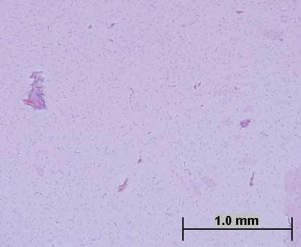
of mononuclear cells surround multifocal blood vessels within
infection. The significance of the intranuclear inclusion bodies
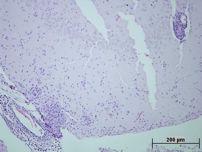
the brainstem, cerebral cortex and meninges (Fig 1c). Focally
within the liver of this possum is uncertain.
CASE HISTORY
the meninges are markedly thickened with a mononuclear cell
A ten year old female brushtail possum (Trichosurus
infiltrate (Fig 1d).
Non-suppurative meningoencephalitis and choroiditis has been
vulpecula) was submitted to a veterinary clinic with a
reported in brushtail possums in Australia since 1985, when
unilateral cataract, ataxia, poor weight and pale gums. The
MORPHOLOGICAL DIAGNOSIS
the clinical syndrome of blindness and ataxia was first
animal was thought to have ingested rodenticides and blood
described as “wobbly possum syndrome”. A similar set of
tests revealed anaemia (PCV 18 L/L). The possum was treated
clinical signs and histological lesions has been identified in
with amoxicillin and vitamin K, and it improved over the
Extensive nonsuppurative meningoencephalitis
brushtail possums in New Zealand, but the relationship
following 2 weeks (the PCV increased to 35L/L). The possum
Focally extensive acute cerebral haemorrhage and malacia
between the two syndromes is uncertain. Although there has
did not eat well, it began loosing weight, and its ataxia
Multifocal eosinophilic intranuclear inclusion bodies - liver
been speculation that “wobbly possum syndrome” in Australia
progressed. The possum became dehydrated (PCV48 L/L,
is likely to be associated with a viral pathogen, no aetiological
GROSS PATHOLOGY
Gross post mortem examination revealed no significant
REFERENCES (Abstracts on file)
findings. Formalin fixed tissues were submitted to the
PERROTT M.R.F. MEERS J. COOKE M.M. WILKS C.R.
(2000) A neurological syndrome in a free-living population of
possums (Trichosurus vulpecula). [Journal article] New
HISTOPATHOLOGY
Zealand Veterinary Journal. 48: 1, 9-15. 16 ref.
Lesions are not evident within the following tissues:
THOMPSON E.G. MCLEOD B.J. GILL J.M. (1999) The
myocardium, ovary, oviducts/vagina, pancreas, spleen.
prevalence of Wobbly Possum disease in a bush/farmland
Liver: Hepatocytes contain small quantities of brown
environment. [Conference paper. Journal article] Proceedings
cytoplasmic pigment. Multifocal hepatocytes contain
of the New Zealand Society of Animal Production. 59: 233-
eosinophilic intranuclear inclusion bodies and have
O'KEEFE J.S. STANISLAWEK W.L. HEATH D.D. (1997)
Kidney: Scattered renal tubules contain small quantities of
Pathological studies of wobbly possum disease in New
mineral or cellular casts. Neutrophils are evident within
Zealand brushtail possums (Trichosurus vulpecula). [Journal
glomeruli. A small number of interstitial lymphoid aggregates
article] Veterinary Record. 141: 9, 226-229. 19 ref.
are evident within the renal interstitium. Eye: The sample is fragmented. The retina is artefactually separated from the choroid. No ganglion cells are evident
within the retina. The lens protein at the lateral aspects of the
lens is liquefied. Lenticular epithelial cells have migrated to a
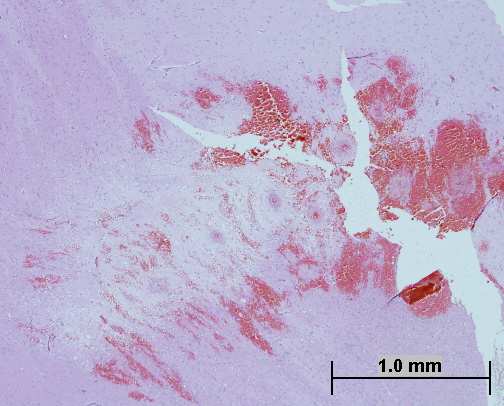
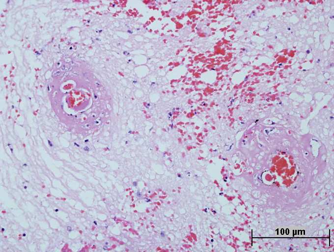
Fig 1. Sections of brain tissue with foci of haemorrhage and
position within the lenticular protein. The connective tissue
malacia (a) and b)), and perivascular mononuclear cell cuffs
surrounding the optic nerve contains a light infiltrate of
(c) and d)). Clefts in the tissues are processing artefacts H & E
mononuclear cells and a small number of neutrophils. The
optic tract itself contains scattered mononuclear cells and 1 - 2
COMMENTS
cell layer perivascular cuffs composed of mononuclear cells.
The lesions in the optic nerve and brain are most suggestive of
Brain: An extensive tract of malacia and haemorrhage is
an acute to subacute viral infection. Eosinophils were not
evident at the junction of the cerebral cortex and the dorsal
evident within the inflammatory infiltrate within the nervous
thalamus (Fig 1a). Blood vessels in this region exhibit
tissue and there is no other evidence of Angiostrongylus sp. or
fibrinoid necrosis (Fig 1b). One to three cell layer thick cuffs
protozoal (Toxoplasma gondii or Neospora caninum)
Case interpretation: Karrie Rose. Case construction: Damien Higgins
SciAnNews Individual Variation and the Acceptance of Average Bioequivalence by Laszlo Endrenyi & Miklos Schulz Drug Information Journal, Vol. 27, pp. 195-201, 1993 0092-8615/93 Printed in the USA. All rights reserved. Copyright (C) 1993 Drug Information Association Inc INDIVIDUAL VARIATION AND THE ACCEPTANCE OF AVERAGE BIOEQUIVALENCE Department of Pharmacology, University of Toro
Bedeutung von Epikutantest und Lymphozytentransformationstest für die Diagnostik von Typ IV-Sensibilisierungen. Stellungnahme des Deutschen Berufsverband der Umweltmediziner Significance of the patch test and the lymphocyte transformation test in the diagnostic of type IV-Statement of the German professional association for enviromental medicine Frank Bartram1, Hans-Peter Donate2, K


 NON-SUPPURATIVE MENINGOENCEPHALITIS IN A BRUSHTAIL POSSUM (Trichosurus vulpecula) (CASE 1411.1)