Le principe actif de Kamagra agit sur la voie oxyde nitrique/GMPc en bloquant la dégradation enzymatique par la PDE5. Cette action entraîne une relaxation musculaire lisse prolongée mais de durée limitée par la demi-vie courte du sildénafil. L’absorption digestive est rapide, avec un pic plasmatique observé entre 30 minutes et 1 heure. Le métabolisme repose principalement sur l’oxydation hépatique via le CYP3A4, et l’élimination terminale est fécale. Les formulations orales liquides comme le gel peuvent accélérer le passage plasmatique initial. Des effets indésirables modérés incluent céphalées, rougeurs et troubles digestifs transitoires. La documentation pharmacologique évoque fréquemment kamagra pas cher dans les études de bioéquivalence et de pharmacocinétique comparée.
Microsoft word - 2_11-19_haider m. hamzah.doc
CONJUGATIVE PLASMID-BORNE ANTIBIOTIC RESISTANCE GENES IN PSEUDOMONAS AEROGENOSA * Haider M. Hamzah, ** Sinaa Muhammed Ali, * Ibrahim Sulaiman Hamad * Dept. of Biology, College of Science, University of Sulaimani, Kurdistan region- ** Dept. of Community health, Technical College, Foundation of Technical Education, Sulaimani, Kurdistan region-IRAQ [email protected] [email protected] Abstract
A total of 6 isolates of P. aeruginosa were isolated from premature unit, oxygen administration room and blood of patients admitted in pediatric hospital in Sulaimani. The isolates were identified as P.aeruginosa according to the diagnostic keys. All isolates were tested for susceptibility towards 10 different antibacterial drugs and they exhibited multiresistant for several choice antibiotics (amikacin, ampicillin, carbencillin, chloramphenicol, cefotaxime, neomycin, and trimethoprim- sulfamethoxazole), with varied susceptible to rifampicin, gentamicin, and tetracycline. Agarose gel electrophoresis showed two different plasmid bands in P. aeruginosa isolate which is resistant to 7 antibiotics. One of these bands was ranged between 41 to 45 kbp and identified as conjugative plasmid and it confirmed through conjugation process with E. coli. Transconjugants were selected on agar plate containing antibiotics (rifampicin, cefotaxime, and ampicillin) and was recovered at frequencies in range of 10-5-10-6 per recipient. Data suggest that bacterial contamination of different units of pediatric hospital should be reduced or eliminated to minimize nosocomial infection risks related to transfer of antibiotic-resistant bacteria to other patients. Key words: Multiple-Antibiotic-Resistant, P. aeruginosa. Conjugative plasmids. ﺎﻴﺭﺘﻜﺒ ﻲﻓ ﻲﻨﺍﺭﺘﻗﻻﺍ ﺩﻴﻤﺯﻼﺒﻟﺍ ﻰﻠﻋ ﺔﻟﻭﻤﺤﻤﻟﺍ ﺔﻴﻭﻴﺤﻟﺍ ﺕﺍﺩﺎﻀﻤﻠﻟ ﺔﻤﻭﺎﻘﻤﻟﺍ ﺕﺎﻨﻴﺠﻟﺍ ﺎﻫﺭﻴﺒﻌﺘﻭ Pseudomonas aeruginosa ﺔﺼﻼﺨﻟﺍ
ﻰﻀﺭﻤﻟ ﻡﺩﻟﺍ ﺝﺫﺎﻤﻨﻭ ﻥﻴﺠﺴﻜﻭﻻﺍ ﺀﺎﻁﻋﺍ ﺔﻓﺭﻏ ،ﺝﺩﺨﻟﺍ ﺓﺩﺤﻭ ﻥﻤ
P. aeruginosa ﺎﻴﺭﺘﻜﺒﻟ ﺔﻟﺯﻋ
ﺢﻴﺘﺎﻔﻤﻟﺍ ﺏﺴﺤ P. aeruginosa ﺎﻬﻨﺍ ﻰﻠﻋ ﺕﻻﺯﻌﻟﺍ ﺕﺼﺨﺸ .ﺔﻴﻨﺎﻤﻴﻠﺴﻟﺍ ﻲﻓ لﺎﻔﻁﻻﺍ ﻰﻔﺸﺘﺴﻤ ﻲﻓ ﻥﻴﻠﺨﺍﺩ ﺽﻌﺒﻟ ﺓﺩﺩﻌﺘﻤ ﺔﻤﻭﺎﻘﻤ ﺕﺭﻬﻅﺍﻭ ﻱﻭﻴﺤ ﺩﺎﻀﻤ 10 ﻩﺎﺠﺘ ﺔﻴﺴﺎﺴﺤﻠﻟ ﺕﻻﺯﻌﻟﺍ ﻊﻴﻤﺠ
ﺕﺭﺒﺘﺨﺍ .ﺔﻴﻔﻴﻨﺼﺘﻟﺍ
,ﻥﻴﺴﻴﺎﻤﻭﻴﻨ ,ﻥﻴﺴﻜﺎﺘﻭﻔﻴﺴ ,لﻭﻜﻴﻨﻴﻔﻤﺍﺭﻭﻠﻜ ,ﻥﻴﻠﺴﻨﺒﺭﺎﻜ ,ﻥﻴﻠﻴﺴﻴﺒﻤﺍ ,
ﻥﻴﺴﺎﻜﻴﻤﺍ ﺔﻴﻭﻴﺤﻟﺍ ﺕﺍﺩﺎﻀﻤﻟﺍ
.ﻥﻴﻠﻜﻴﺎﺴﺍﺭﺘﻴﺘ ﻭ،ﻥﻴﺴﺒﻤﺎﻔﻴﺭ ,ﻥﻴﺴﻴﺎﻤﺘﻨﺠﻟﺍ ﻩﺎﺠﺘ ﺔﺘﻭﺎﻔﺘﻤ ﺔﻴﺴﺎﺴﺤ ﻊﻤ ,(لﻭﺯﺎﺴﻜﻭﺜﻴﻤﺎﻔﻟﻭﺴ
H. M. Hamzah, S. M. Ali, I. S. Hamad CONJUGATIVE PLASMID.
ﺔﻤﻭﺎﻘﻤﻟﺍ P. aeruginosa ﺔﻟﺯﻌﻟﺍ ﻲﻓ ﺔﻴﺩﻴﻤﺯﻼﺒ ﻥﻴﺘﻤﺯﺤ ﺩﻭﺠﻭ ﺯﻭﺭﺎﻜﻻﺍ ﻡﻼﻬﺒ ﻲﺌﺎﺒﺭﻬﻜﻟﺍ ﻥﻼﺤﺭﻟﺍ ﺭﻬﻅﺍ ﺩﻴﻤﺯﻼﺒﻜ ﺹﺨﺸ ﻥﻴﺘﻤﺯﺤﻟﺍ ﻥﻤ ﺩﺤﺍﻭ ،ﺱﻴﺒ ﻭﻠﻴﻜ
41 ﻥﻴﺒﺎﻤ ﺎﻬﻤﺠﺤ ﺡﻭﺍﺭﺘ .ﺔﻴﻭﻴﺤ ﺕﺍﺩﺎﻀﻤ ﺔﻌﺒﺴﻟ
ﻱﻭﺘﺤﻤﻟﺍ ﺏﻠﺼﻟﺍ ﻁﺴﻭﻟﺍ ﻰﻠﻋ ﺕﺎﻨﺭﺘﻘﻤﻟﺍ ﺀﺎﻘﺘﻨﺍ ﻡﺘ .E. coli ﺎﻴﺭﺘﻜﺒ ﻊﻤ ﻥﺍﺭﺘﻗﻻﺍ ﺔﻴﻠﻤﻋ لﻼﺨ ﻥﻤ ﺩﻜﺄﺘﻭ ﻥﺭﺘﻘﻤ
10 ﺩﻭﺩﺤﺒ ﺩﺩﺭﺘﻟﺍ لﺩﻌﻤ ﻥﺎﻜﻭ (ﻥﻴﻠﺴﻴﺒﻤﺍﻭ ،ﻡﻴﺴﻜﺎﺘﺎﻔﻴﺴ ،ﻥﻴﺴﺒﻤﺎﻓﺭ ) ﺔﻴﻭﻴﺤﻟﺍ ﺕﺍﺩﺎﻀﻤﻟﺍ ﻰﻠﻋ
ﻥﻤ لﻴﻠﻘﺘﻠﻟ لﺍﺯﻴ ﻭﺍ ﺽﻔﺨﻴ ﻥﺍ ﺏﺠﻴ لﺎﻔﻁﻻﺍ ﻰﻔﺸﺘﺴﻤ ﺕﺍﺩﺤﻭ ﻑﻠﺘﺨﻤﻟ ﻱﺭﻴﺘﻜﺒﻟﺍ ﺙﻭﻠﺘﻟﺍ ﻥﺎﺒ ﺞﺌﺎﺘﻨﻟﺍ ﺕﺤﺭﺘﻗﺍ
.ﻥﻴﺭﺨﺍ ﻰﻀﺭﻤﻟ ﺔﻴﻭﻴﺤﻟﺍ ﺕﺍﺩﺎﻀﻤﻠﻟ ﺔﻤﻭﺎﻘﻤﻟﺍ ﺎﻴﺭﻴﺘﻜﺒﻟﺍ لﺎﻘﺘﻨﺎﺒ ﺔﻗﻼﻌﻟﺍ ﺕﺍﺫ ﺕﺎﻴﻔﺸﺘﺴﻤﻟﺍ ﻯﻭﺩﻋ ﺭﺎﻁﺨﺍ
Introduction Nosocomial infections (hospital acquired infection) are infections not present or incubating before hospital admission, and continue to complicate the clinical course of critically ill patients and consequently to create substantial economic and human costs [1]. Nosocomial infection typically affect patients who are immunocompromised because of ages underlying disease or medical or surgical treatments, also nosocomial infection rates in adult and pediatric intensive care units or approximately three times higher than elsewhere in hospitals [2]. In general, it appeared that the majority of hospitalized patients are infected by Gram negative multiresistant bacteria such as Klebsiella spp., Enterobacter spp., Pseudomonas spp., Acinetobacter spp., Serratia spp [3]. It was reported that in hospitals sinks, respiratory therapy equipments and antiseptic or detergent solution can act as reservoir of Pseudomonas aeruginosa and cross transmission may occur via the hands of the health care staff or through contaminated materials or reagents [4].The extensive use of antibiotics in clinical therapy of human infectious diseases during the past 50 years has resulted in the emergence and rapid global spread of antibiotic resistance determinants [5, 6]. These determinants are usually located on mobile genetic elements such as conjugative plasmids [7] or conjugative transposons [8], which ensure their dissemination among bacterial populations via horizontal gene transfer (HGT). As a consequence of the lateral transfer of resistance genes, multiply resistant human pathogens, such as Shigelladysenteriae and Pseudomonas aeruginosa, have emerged, which meanwhile pose serious health problems [9]. The obvious importance of conjugative antibiotic resistance plasmids for human and animal health has raised the issue of the incidence of such plasmids in the environment. The main objective of this study was to isolation of conjugative plasmid harboring antibiotic resistance genes from Pseudomonas aeruginosa strains isolated from premature unit, oxygen administration room and blood of patients admitted in pediatric hospital in Sulaimani. Materials and Methods
1- Isolation and Identification of P. aeruginosa The strains of Pseudomonas aeruginosa were isolated from the premature unit, oxygen administration room and blood of patients (Pediatric hospital in Sulaimani) by emerging sterile swabs on the surface of area tested (e.g. physician hands, binasal
cannula, sucker) and directly transferred to laboratory for culturing on blood and nutrient agar plates as well as on selective medium (cetrimide agar) [10]. Plates were incubated at 37 °C for 18-24 hrs. Several biochemical tests were done to differentiate P. aeruginosa from other species. These include: IMViC test, oxidase test, urease test, H2S production, and sugar fermentation (mannitol, glucose, and lactose).
2- Antibiotic Susceptibility Test The antibiogram test using Kirby-Bauer method was carried out using Mueller-Hinton agar plates [11]. Bacterial isolates were grown in nutrient broth for 24 h and the broth swabbed evenly onto the surface of Mueller-Hinton agar plates using sterile cotton swabs and the covered plates allowed drying. Antibiotic-impregnated filter paper discs were placed on the surface of the agar and the plates were incubated at 35 °C for 24 h. Inhibition zones were then measured to the nearest millimeter. Inhibition zones were indicated by a lack of bacterial growth due to inhibitory concentrations of antibiotics diffused into semisolid culture media (agar) beneath the antibiotic- impregnated disc. Antibiotic and their doses tested were as follows: Amikacin (AMI, 30 µg), ampicillin (AP, 10 µg), carbencillin (CAR, 100 µg), chloramphenicol (C, 30 µg), cefotaxime (CTX, 30 µg), rifampicin (RF, 30 µg), tetracycline (TE, 30 µg), gentamycin (CN, 10 µg), neomycin (N, 30 µg), and trimethoprim-sulfamethoxazole (SXT, 1.25 µg). Antibiotic resistance was determined by comparing bacterial isolate inhibition zone diameters to established values [11]. 3- Analysis of plasmid content Alkaline lysis method [12] was used to extract plasmids. Single colony of the bacterial isolate was grown in 10 ml of Luria Bertain (LB) broth containing 50 mg/ml ampicillin and incubated at 37 °C for 24 hr with shaking. Bacterial cells were harvested by centrifugation at 10000 rpm for 10 min. The pellet was resuspended in 10 ml of EST/saline buffer. The suspension centrifuged at 10000 rpm for 10min (the step 2, 3 repeated twice). The pellet was resuspended in 0.2 ml of solution I (prepared by mixing 50 mM glucose, 25 mM Tris-Cl, pH 8 and 10 mM EDTA, pH 8, the solution was prepared in batches of 100 ml then the solution was sterilized by autoclave and stored at 4°C), then transferred to sterile eppendorf tube and omitted for 5 minutes at room temperature. Aliquot of 0.4 ml of solution II (prepared by mixing 0.2 N NaOH, freshly diluted from 10 N stock, and 1% SDS) was added to the mixture and then the eppendorf tube was gently inverted for many times and omitted in ice bath for 15 min. Aliquot of 0.3 ml of cold solution III (3 M sodium acetate, pH 4.8, it was prepared by mixing 60 ml of 5 M potassium acetate, 11.5 ml glacial acetic acid and 28.5 ml of distilled water) was added to the mixture above and the eppendorf tube was inverted for many times before putting on ice bath for 5 min. The mixture was centrifuged at 14000 rpm for 5 min. Aliquot of 0.5 ml of the supernatant was transferred to the new sterile eppendorf, then an equal volume of phenol-chloroform- isopropanol solution was added and mixed very well then the solution objected to centrifugation as previous step. The upper layer was transferred to the new sterile eppendorf, and both the middle and lower layer omitted, this step was repeated till the protein was excluded from the solution. Aliquot of 3 M of cold sodium acetate in ratio of 0.1 volume was added to the supernatant that contain plasmid DNA and mixed very well. Double volume of cold absolute ethanol was added to the suspension and mixed gently and then left at -10 °C for two hrs. The mixture was H. M. Hamzah, S. M. Ali, I. S. Hamad CONJUGATIVE PLASMID.
centrifuged at 12000 rpm for 15 min then the ethanol was excluded from the solution, and the pellet was washed by 70% ethanol and then centrifuged as in the previous step. The eppendorf was inverted on sterile filter paper and the pellet dissolved in 50µl of TE buffer and stored at -20°C [12]. 4- Bacterial conjugation The bacterial conjugation was achieved according to [13]. A single colony of donor strain was inoculated into 50 ml LB broth containing appropriated antibiotic. A single colony of E.coli MM294 rifr (act as recipient cells) was inoculated into 50 ml LB broth containing 100 µg/ml of rifampicin. The cultures were incubated overnight at 37°C for 18 hr with vigorous shaking until to obtain heavy growth (O.D. 0.5). About 5×10⁷ of Pseudomonas aeruginosa donor cell and 1×10⁸ recipients were mixed in sterile eppendorf. The pellet was resuspended in 50µl of LB and the cells were transferred to 0.22 µm millipore filter on LB agar plate. The LB agar plates which contain millipore filter were incubated at 37°C for 1-2 hr. The cells were resuspended by placing filter in a tube containing 0.5 ml of 0.85% saline and agitating the tube on a vortex. Serial dilutions (10-1-10-8) were prepared of the mating mixture. Aliquot of 0.1 ml of dilution (10-1 to 10-6) was spread on LB agar containing rifampcin (100µl/ml), cefotaxime (50 µl/ml), and ampicillin (150 µl/ml) with rifampicin. Aliquot of 0.1ml of original overnight culture of donor and recipient cells was spread on the same medium to determine the frequency of antibiotic resistance spontaneous mutation, 0.1 ml of dilution 10-6 and 10-8 was spread on LB agar containing rifampcin (100 µl/ml) to estimate the number of recipient cells in the mating. All plates were incubated at 37°C for overnight. The control treatment was included aliquot of 0.1 ml of donor cells was transferred to rifampcin plate. Aliquot of 0.1 ml of recipient cell E. coli MM294 rifr were transferred to plates containing antibiotic of donor strain. All plates were incubated at 37°C for overnight. The colonies that had grown on the selection plate were counted and the number of bacteria per ml in mating mixture exhibiting the phenotype rifampicin (total recipient) and rifr, ampr (transconjugant) was calculated [13]. Results and Discussion
1- Isolation and Identification of P. aeruginosa Several studies have demonstrated that conjugative antibiotic resistance plasmids are widespread in nature. Such plasmids have been identified in habitats including the marine environment [14], river and lake water [15], as well as in raw and treated sewage [16]. A total of 121 samples were collected from premature unit, oxygen administration room and blood of patients in the pediatric hospital-Sulaimani. Table 1 shows the samples obtained from different sources and site of the hospital. Table (1): Site, number and samples of isolates of P. aeruginosa. Site of isolation isolates Premature unit Oxygen administration room Patient sample Total of samples
Only 6 isolates of P. aeruginosa were detected particularly from patient's samples, sucker, binasal cannula and oxygen administration. The characteristics of P. aeruginosa strain are summarized in Table 2. It was identified as P. aeruginosa according to Bergey's Manual of Systematic Bacteriology [17].
Table (2): Characteristics of local isolate of Pseudomonas aeruginosa. Characteristic Note: D, different strains gave different results.
P. aeruginosa is important and frequent nosocomial pathogens indeed in hospitals, sink, respiratory therapy equipment and antiseptic or detergent solutions can acts as reservoirs of this organism which is responsible for a wide range of hospital acquired infection and bacteremia, outbreaks of nosocomial infection due to P. aeruginosa have been reported especially in ICU[4]. 2- Antibiotic Susceptibility Testing Refer to Table 3 for a summary of P. aeruginosa isolates antibiotic resistance. Six isolates were resistant to 7 antibiotic groupings that included amikacin, ampicillin, carbencillin, chloramphenicol, cefotaxime, neomycin, and trimethoprim- H. M. Hamzah, S. M. Ali, I. S. Hamad CONJUGATIVE PLASMID.
sulfamethoxazole. Four bacterial isolates were resistant to gentamicin; and one isolate was resistant to rifampicin, another one resistant to tetracycline.
Table (3): Multiple-antibiotic resistant isolates of Pseudomonas aeruginosa. Antibiotic Total multiple- isolates resistant sensitive antibiotic resistant isolates isolates isolates (%)
It is well known that P. aeruginosa is the major problem with respect to antibiotic susceptibility [18]. It was found that multidrug resistant of P. aeruginosa nsocomial infections are increasingly recognized world wide, the existence of metallo-β- lactamase and extended spectrum β-lactamase producing isolates exhibiting resistance to most β-lactam antibiotics [19]. Cells resistance in P. aeruginosa may develop rapidly and by multiple mechanisms including production of specific enzymes, outer membrane permeability changes and active efflux system [20]. It was reported that since 1998, P. aeruginosa isolates in Brazil were resistant to all commercially available antimicrobial agents [21], while another study revealed that all the P. aeruginosa isolates are insensitive to all potentially active antibiotics except colistin [22]. 3- Plasmids in P. aeruginosa Plasmid may reside in cells either as a single copy or as multiple copies, large plasmid tends to have a lower copy number, whereas smaller plasmid may be present in more than 20 copies per cell and several studies have demonstrated that conjugative antibiotic resistance plasmids are widespread in nature [23]. Resistance in P. aeruginosa may be chromosomal born and in general, clinical strains may harbor multiple plasmids. Plasmids harboring in P. aeruginosa were equally well resolved by alkaline procedure (Fig.1). A total of two different plasmid bands were identified in P. aeruginosa, one of them with 40-45 kb in sizeappeared in well No. 1. The plasmid sizes calculated on the basis of that found in E. coli containing R 751 plasmid (well No. 2) which range its size about 51 kb. Another smaller unidentified plasmid appeared in P. aeruginosa isolate but its appeared as non conjugative plasmid. Rice et al (1993), reported that some plasmid exceed 180 kb in size and encode for a wide varieties of antimicrobial resistance genes [24]. Fig 1: Plasmids profile in P. aeruginosa isolated from pediatric hospital: (1) P. aeruginosa plasmid contents, (2) E. coli containing R 751 plasmid, (3) Transconjugant E. coli, (4) E. coli MM
To identify distinct conjugative plasmids, transconjugants were first grouped according to their antibiotic resistance patterns. For this purpose, transconjugants grown on the primary selective agar plates were spread on LB medium containing rifampicin, cefotaxime, and ampicillin. P. aeruginosa transconjugants were also tested on LB agar plates containing mixed of the same mentioned antibiotics, and antibiotic resistant colonies arose at frequencies in the range of 10-5 to 10-6 per recipient cell, thus confirming the high selectivity of the selection system. Conclusions Results of this study indicating that the isolates were multiple antibiotic resistant (MAR) are of some importance. MAR of potentially pathogenic bacteria is of public health concern because of the difficulty in treating infections caused by multiple antibiotic resistance bacteria. Treatment of MAR infections often requires much more expensive antibiotics and long-term therapy, substantially increasing the cost of treatment. References 1. Gholam, H.S.B., Ashraf, K.N., Mohammad, R.H., Mohammad, H.G., Parvin, R.F., Mahmood, R.M., Shahla, M. and Alireza, F. (2003). Anti-Pseudomona and anti- Bacilli activity of some medicinal plants of Iran. Daru. 11(4): 157-163. 2. Murray, P.R., Drew, W.L., Kobayashi, G.S. and Thompson, J.H. (1990). Medical Microbiology, Mosby Co., Philadelphia, pp. 119-126. 3. Haley, R.W., Culver, D.H. and White, J.W. (1985). The nation wide nosocomial infection rate: a new need for vital statistic. Am. J. Epidemiol. 121: 159-167. H. M. Hamzah, S. M. Ali, I. S. Hamad CONJUGATIVE PLASMID.
4. Fridkin, S. K., Welbel, S.F. and Weinstein, R.A. (1997). Magnitude and prevention of nosocomial infections in the intensive care unit. Am. Infect. Dis. Clin. North. 11: 479-496. 5. Davies J (1994). Inactivation of antibiotics and the dissemination of resistance genes. Science 264: 375-381. 6. Baquero F, Blazquez J (1997). Evolution of antibiotic resistance. Trends Ecol Evol 12: 482-487. 7. Datta N, Hughes VM (1983). Plasmids of the same Inc groups in Enterobacteria before and after the medical use of antibiotics. Nature 306: 616-617. 8. Saylers AA, Shoemaker NB (1994). Broad host range gene transfer: plasmids and conjugative transposons. FEMS Microbiol Ecol 15: 15-22. 9. Cohen ML (1992). Epidemiology of drug resistance: implications for a post-antimicrobial era. Science 257: 1050-1055. 10. Cheesbrough, M. (1991). Medical laboratory manual for tropical countries. Vol. 2 ELBS. University press, Cambridge, Great Britain, pp.265. 11. Bauer, A.W., Kirby, J.S. and Turck, M. (1966). Antibiotic susceptibility testing by a standardized disc method. Am. J. Clin. Pathol. 45: 493-496. 12. Sambrook, J.; Fritgah, E. and Maniatis, T. (1989). .Molecular cloning: a laboratory manual. Cold spring Harbour laboratory. New York. 13. Connell, O. U. (1984). Genetic transfer in prokaryotes: transformation, transduction, and conjugation. In Advanced molecular genetics edited by Auhler, A and Timmis, K. Springer Verlage, Berline. 14. Sandaa RA, Torsvik VL, Goksùyr J (1992). Transferable drug resistance in bacteria from fish-farm sediments. Can J Microbiol. 38: 1061-1065. 15. Arvanitidou M, Tsakris A, Constantinidis T.C, and Katsouyannopoulos V.C. (1997). Transferable antibiotic resistance among Salmonella strains isolated from surface waters. Wat Res 31:1112-1116. 16. Blazquez J, Navas A, Gonzalo P, Martinez JL, Baquero F (1996). Spread and evolution of natural plasmids harboring transposon Tn5. FEMS Microbiol Ecol 19: 63-71. 17. Krieg, N.R. and Hoh, J.G. (1984). Gram-negative aerobic rods and cocci. In: Bergey's manual of systematic bacteriology, vol.1. (Murray, R.E.G., Brenner, D.J., Bryant, M.P., Hoh, J.G., Krieg, N.R., Moulder, J.W., Sneath, P.H.A. and Staley, J.T, eds). Williams and Wilkines. Baltimore. London, pp. 18. Kremery, V. and Trupl, J. (1994). Nosocomial outbreak of meropenem resistant P. aeruginosa infections in a cancer center. J. Hosp. Infect. 26: 69-71.
19. Luzzaro, F., Mantenooli, E., Perills, M., Lombardi, G., Orlandi, V., Orsatti, A., Amicosante, G., Rossolini, G. and Toniolo, A. (2001). Dynamics of a nosocomial out break of multidrug resistant Pseudomonas aeruginosa producing the PER-Extended spectrum β-lactamase. J. Clin. Microbiol. 39(5):1865-1870. 20. Zarifi, I.Z., Llanes, C., Hler, T., Pechere, J.C. and Plesiat, P. (1999). In vivo emergence of multidrug resistant mutant of Pseudomonas aeruginosa over expressing the active efflux system Mex A, Mex B-OprM. A.A.C. 43(2): 287-291. 21. Pellegrino, F.L.P.C., Teixeira, L.M., Carvalho, M.G.S., Nouer, S.A., Oliveia, M.P., Sampaio, J.I.M., Freitas, A.D., Ferreira, A.L.P., Amorim, E.I.T., Riley, L.W. and Moreira, B.M. (2002). Occurrence of a multidrug resistant Pseudomonas aeruginosa clone in different hospitals in Rio de Janeiro, Brazil. J. Clin. Microbiol. 40(7): 2420-2424. 22. Dubois, V., Arpin, C., Melon, M., Melon, B., Ander, C., Frigo, C. and Quentintune, C. (2001). Nosocomial outbreak due to a multiresistant strain of Pseudomonas aeruginosa P12: Efficacy of cepepim, amikacin therapy and analysis of β-lactam resistance. J. Clin. Microbiol. 39(6): 2072-2078. 23. Droge, M., Puhler, A. and Selbitschka, W. (2000). Phenotypic and molecular characterization of conjugative antibiotic resistance plasmids isolated from bacterial communities of activated sludge. Mol. Gen. Genet. 263: 471-482. 24. Rice, L.B., Willy, S.H., Papanicolaou, G.A. and Jacoby, G.A. (1993). Sequences of MGH-1, YOU-1 and YOU-2 extended spectrum β-lactamases genes. A.A.C.37:2760-2761.
INFORMATION AND CONSENT FORM following braces removal for the office of Dr Kenmuir OVERVIEW This tooth bleaching/whitening technique uses a custom-fitted retainer and peroxide- containing whitening agents. Night wear uses one application at bedtime. Treatment time is generally 2-6 weeks. The more times or longer the solution is applied, the faster the bleaching occurs. However, faster blea
504-456-8515 FLU-MIST IMMUNIZATION 2013/2014 Flu MIST Influenza (flu) is a respiratory disease caused by influenza virus infection. The types, or strains, of influenza virus that cause illness may change from year to year, or even within the same year. People who get flu may have fever, chills, headache, dry cough, and muscle aches, and may be sick for several days to a wee

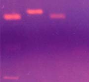
 Fig 1: Plasmids profile in P. aeruginosa isolated from pediatric hospital: (1) P. aeruginosa