Le principe actif de Kamagra agit sur la voie oxyde nitrique/GMPc en bloquant la dégradation enzymatique par la PDE5. Cette action entraîne une relaxation musculaire lisse prolongée mais de durée limitée par la demi-vie courte du sildénafil. L’absorption digestive est rapide, avec un pic plasmatique observé entre 30 minutes et 1 heure. Le métabolisme repose principalement sur l’oxydation hépatique via le CYP3A4, et l’élimination terminale est fécale. Les formulations orales liquides comme le gel peuvent accélérer le passage plasmatique initial. Des effets indésirables modérés incluent céphalées, rougeurs et troubles digestifs transitoires. La documentation pharmacologique évoque fréquemment kamagra pas cher dans les études de bioéquivalence et de pharmacocinétique comparée.
Hercules_06_abstract.dvi
Anomalous Diffraction and Diffraction Anomalous Fine Structure to
study heterostructures and nanostructures
1 Commissariat à l’Energie Atomique, Département de Recherche Fondamentale sur la Matière Condensée,
SP2M/Nanostructure et Rayonnement Synchrotron, 17 rue des Martyrs, 38054 Grenoble Cedex 9, France.
2Université Joseph Fourier, BP 53, F-38041, Grenoble Cedex 09, France. Introduction
The knowledge of strain, vertical and lateral chemical compositions, inter-mixing at the interfaces, i.e. structural prop-
erties at the long and short range order scale, are of great importance to understand the growth mechanism as well as
the electronic and optical properties of the heterostructures and nanostructures. X-ray diffraction is known to be very
powerful for measuring strain fields and correlations. Chemical sensitivity can be obtained using anomalous diffraction
and the local environment of atoms located in an iso-strain region of the nanostructure can be obtained with Diffraction
Anomalous Fine Structure. On the other hand x-ray diffraction is a non destructive method that averages over many
individual nanostructures and gives statistically relevant structural properties. Since thin films or nano-objects grown
onto a bulk substrate have very small scattering volumes, the diffuse scattering from defects in the substrate or thermal
diffuse scattering overwhelm the nanostructures signal. A way the overcome the problem is to perform the experiments
in grazing incidence to reduce the substrate contribution. For more details on the structural properties of self-organised
semiconductor nanostructures one should recommend the reading of a recent review written by Stangl. et al. [1] and refer
to the talk given by T.H. Metzger entitled “X-ray reflectivity and diffraction of nanosystems”.
These x-ray structural studies are a perfect example of the need of high brillance and tunable energy, these unique
capabilities can only be found at a synchrotron radiation facility. Complementary techniques are being used :
1. GISAXS (Grazing Incidence Small Angle X-ray Scattering) for studying morphology, size and position correla-
tions. It can be used to study islands either free standing onto a substrate or buried. Measurements can also be
performed in-situ, under ultra high vacuum, during deposition, to study the growth mechanism, the ripening and the
self organisation of nanostructures [2].
2. X-ray reflectivity to investigate the morphology of surfaces and interfaces as well as the vertical position correlations
3. GIDAFS (Grazing Incidence Diffraction Anomalous Fine Structure) spectroscopy, including Grazing Incidence
GISAXS and x-ray reflectivity are rather well established and quantitative techniques (regarding GISAXS see for instance
ref. [3]). in the following sections we give more details about Grazing Incidence Anomalous Diffraction and GIDAFS
Diffraction Anomalous Fine Structure
Diffraction Anomalous Fine Structure (DAFS) spectroscopy is based on resonant elastic x-rays scattering. A DAFS
experiment is the measurement of the elastic scattering intensity as a continuous function of the incoming x-ray beam
energy in the vicinity of absorption edges. It provides information about the chemical state and the local environment
of the resonant atom (also known as the anomalous atom), like x-ray Absorption Fine Structure (XAFS) spectroscopy.
But in contrast to XAFS, it is a chemical-selective and site-selective spectroscopy. Like Multiple-wavelength Anomalous
Diffraction (MAD), DAFS provides crystallographic phases and structure factor amplitudes that give information on the
long-range crystallographic structure (see for instance the review by Hodeau et al. [4]). The technique is being developped
since several years at beamline BM2-D2AM at the ESRF [5].
The DAFS spectroscopy has proven to be a very successful tool to study the crystallographic structure of thin films,
superlattices and interfaces. As a matter of fact anomalous scattering gives pertinent diffraction data that are very useful to
recover composition gradients and atomic displacements at the interfaces [6]. On the other hand the diffraction oscillations
are used to recover 3D local structure of sites or iso-strain regions selected by the diffraction condition. In this sense we
do believe that DAFS it is not yet exploited by the scientific community as it should be, according to its great capabilities.
For instance, in case of disordered samples, slightly different local environments coexist and it is well known that XAFS
spectroscopy fails to give pertinent information or at least the XAFS data are difficult to analyze. One should note
that, although the DAFS data contain the contributions of both the real and imaginary parts of the complex anomalous
scattering factors (XAFS data is proportional to the imaginary part), they can be analyze, in the extended region, like the
Extended-XAFS data by using the same analysis packages [7]. Also, efficient program for simulating the near edge DAFS
Grazing Incidence Anomalous Diffraction
Grazing anomalous diffraction consists in measuring diffraction curve at several energies in the vicinity of the absorption
edge of one sample’s element. By tuning the energy only the scattering power of the element is changed and the sample
compostion can be obtained from intensity ratio. Combined with the “iso-strain scattering” method [9], anomalous scat-
tering at the Ge K-edge was used to recover the out of plane Ge composition gradient in uncovered Ge/Si(001) islands
[10, 11]. Anomalous diffraction was also used to investigate the lateral composition of uncapped Ge domes grown on
Si [12]. This method is suitable for uncapped islands with a strong lattice mismatch. As a matter of fact, to be suitable
for devices, the nanostructures are encapsulated or embedded in a superlattice, they must be homogeneous in size, shape
and composition, to provide well defined emission wavelengths. We have used Multiwavelenght Anomalous Diffraction
(MAD) in grazing incidence diffraction to study the strain, size and composition of InAs Quantum Sticks, grown on InP
and covered with a 10 nm thick InP cap layer. The partial structure factor FAs (i.e. the Fourier Transform) of all As
atoms, was directly extracted, allowing to obtain the average height and strain of the InAs QSs and to determine their
composition and check the As/P exchange [13]. DAFS oscillations, in the energy range above the As edge, can also be
used to obtain direct information on the local composition and on strain accomodation inside the sticks (see section 4)
Grazing Incidence Diffraction Anomalous Fine Structure
Since only recently DAFS in grazing incidence (GI-DAFS) is being developped at the beamline BM2-D2AM at the ESRF.
It has been used, for the first time, to study uncovered InAs quantum wires (QWrs) grown on InP(001) [14]. The QWrs
10] direction with a typical length above 5 µm, a height between 0.6 and 2 nm, a period of 20
nm with an equivalent InAs coverage of about 2.5 monolayers (fig. 1a). Grazing Incidence DAFS spectra were mesured
at the maximum of the QWrs correlation satellites, near the in-plane (420) and (440) InP substrate Bragg peaks at the As
Extended DAFS oscillations that appear after the edge (fig. 1b-a) were analysed according to an EXAFS data process-
ing scheme to get local parameters such as distances and atomic populations. The polarisation of the incoming photons
was perpendicular to the surface, so the As and P next nearest neighbour atoms contribution to EDAFS is due only to the
Figure 1: (a) AFM tridimensional view of InAs quantum wires on InP buffer ; (b-a) InAs quantum wire Grazing IncidenceExtended DAFS oscillations, after background subtraction, with best fit (continuous line), (b-b) EXAFS of the quantumwires. The curves have been rescaled for clarity ; (c-a) FT of quantum wire EDAFS, with best fit (continuous curve), (c-b)FT of quantum wire EXAFS. The curves have been rescaled for clarity.
out-of-plane atoms. The relevant results are the As-P distance, found at 4.17 Å, close to the P-P distance in bulk InP (4.15
Å), therefore, we could exclude the hypothesis of a fully relaxed InAsP epilayer. The P atoms contributing to EDAFS
belong to the interface region, 0.5-2 monolayers, and the core of the quantum wire is essentially strained InAs.
For comparison we measured a glancing-angle EXAFS spectrum at the As K-edge (fig. 1b-b) at beamline BM8
(GILDA, ESRF). The spectrum shows a clear As oxide shape with a strong low-frequency component that corresponds to
a strong peak at 1.2 Å in its Fourier Transform (FT) (fig. 1c-b). The oxide layer causes a significant loss of information
in particular for shells beyond the first one, whereas, for a DAFS spectrum, it lowers the overall diffracted intensity and
the jump at the edge but it does not perturb the fine structure signal of the interesting atoms.
At present, we are still working intensively on the developpment of both the GIDAFS experiment at beamline BM2
(ESRF) and the data analysis. The method is applied to the study of the GaN/AlN and Ge/Si quantum dots to recover
strain fields and composition gradients [15, 16, 17]. Polarisation dependence
As for XAFS, DAFS spectra depend on the x-ray beam polarization direction. For x-ray absorption, the crystallographic
point group governs that dependence. For instance the absorption is isotropic for a cubic point group even if the site
symmetry of the absorber is not [18]. In case of DAFS instead, the situation is quite different. The polarization of
the incoming and outgoing x-ray beams must be taken into account. Via virtual mutipole transitions (mainly dipolar,
quadrupolar or an interference of both), the energy dependences in the σ − σ and σ − π channels upon the incomingx-ray beam energy as well as upon the azimuthal angle (corresponding to a rotation about the scattering vector) reveal the
Anisotropy of the Susceptibility Tensor (ATS) [19, 20], i.e. the site symmetry of the resonant atom.
As far as structural properties are concerned, the polarization dependence may be used as in x-ray absorption. For
instance, in strained thin films or superlattices of materials which are cubic in the bulk, the interfaces may be non cubic
at the atomic scale. It is a fact that distortions in strained semiconductors are rather small. With the XAFS or GIDAFS
spectroscopies these distortions are to be evaluated with the second nearest neighbors, and it is often difficult to obtain
good results with data obtained with either the polarization in the plane or perpendicular. It is very efficient to perform a
co-refinement of 2 spectra obtained with both polarizations. In that case, one should measure GIDAFS spectra with the
polarization of the incoming beam in and out of the growth plane to probe in- and out of plane local distortions. Conclusion
In conclusion, the grazing incidence anomalous diffraction and GI-DAFS are powerful tools to study nanostructures. Its
main interest is to provide structural properties (strain accomodation and composition) inside “iso-strain” region that are
selected by the diffraction condition. The ultimate goal is to map these properties in the three dimension, to get detailed
information on lateral composition gradients. Therefore it is of a great importance to be able to carry out anomalous
diffraction 2D mappings and GI-DAFS measurements together on a dedicated beamline with a high brillance and high
beam position and energy stabilities. We would also like to stress that DAFS can be regarded as the meeting point of the
diffraction and absorption scientific communities with a potentially great scientific impact. References
[1] J. Stangl, V. Holy, and G. Bauer. Structural properties of self-organized semiconductor nanostructures. Rev. Mod.
[2] G. Renaud, R. Lazzari, C. Revenant, A. Barbier, M. Noblet, O. Ulrich, F. Leroy, J. Jupille, Y. Borensztein, C.R.
Henry, J.P. Deville, F. Scheurer, J. Mane-Mane, and O. Fruchart. Real-time monitoring of growing nanoparticules.
[3] C. Revenant, F. Leroy, R. Lazarri, G. Renaud, and C.R. Henry. Quantitative analysis of grazing incidence small-angle
x-ray scattering : Pd/mgo(001) growth. Phys. Rev. B, 69(035411), 2004.
[4] J.L. Hodeau, V. Favre-Nicolin, S. Bos, H. Renevier, J.E. Lorenzo, and J.F. Bérar. Resonant diffraction. Chem. Rev.,
[5] H. Renevier, S Grenier, S Arnaud, J.F. Bérar, B. Caillot, J.L. Hodeau, J.P. Simon, A. Letoublon, M.G. Proietti, and
B. Ravel. Diffraction anomalous fine structure spectroscopy at beamline bm2 at the european synchrotron radiation
facility. J. of Synchrotron Rad., 10:435–444, 2003.
[6] H. Renevier, J.L. Hodeau, P. Wolfers, S. Andrieu, J. Weigelt, and R. Frahm. Selective study of fe atoms at the
interfaces of an Fe/Ir(100) superlattice by means of diffraction anomalous fine structure. Phys. Rev. Lett., 78:2775–
[7] M.G. Proietti, H. Renevier, J.L. Hodeau, J Garcia, J.F Bérar, and P. Wolfers. Diffraction-anomalous-fine-structure
spectroscopy applied to the study of III-V strained semiconductors. Phys. Rev. B, 59:5479–5492, 1999.
[8] Y. Joly. X-ray absorption near-edge structure calculations beyond the muffin-tin approximation. Phys. Rev. B, 63,
[9] I. Kegel, T.H. Metzger, A. Lorke, J. Peisl, J. Stangl, G. Bauer, K. Nordlund, W.W. Schoenfeld, and P.M. Petroff.
Determination of strain fields and composition of self-organized quantum dots using x-ray diffraction. Phys. Rev. B,
[10] R. Magalhaes-Panagio, G. Medeiros-Riberio, A. Malachias, S. Kycia, T.L. Kamins, and R. Stan. Direct evaluation
of composition profile, strain relaxation, and elastic energy of ge:si(001) self-assembled islands by anomalous x-ray
scattering. Phys. Rev. B, 66(6):245312–245320, 2002.
[11] T.U. Schülli, J. Stangl, Z. Zhong, R.T. Lechner, M. Sztucki, T.H. Metzger, and G. Bauer. Direct determination of
strain and composition profiles in sige islands by anomalous x-ray diffraction at high momentum transfer. Phys. Rev.Lett., 90:66105–66109, 2003.
[12] A. Malachias, S. Kycia, G. Medeiros-Riberio, R. Magalhaes-Panagio, T.L. Kamins, and R.S. Williams. 3d composi-
tion of epitaxial nanocrystals by anomalous x-ray diffraction : observation of a si-rich core in ge-domes on si(001). Phys. Rev. Lett., 91:176101, 2003.
[13] A. Letoublon, V. Favre-Nicolin, H. Renevier, M.G. Proietti, C. Monat, M. Gendry, O. Marty, and Priester C. Strain,
size and composition of inas quantum sticks, embedded in inp, determined via grazing incidence x-ray anomalous
diffraction. Phys. Rev. Lett., 92:186101, 2004.
[14] S. Grenier, M.G. Proietti, H. Renevier, L. Gonzalez, J.M. Garcia, and J. Garcia. Grazing-incidence diffraction
anomalous fine structure of inas/inp(001) self-assembled quantum wires. Europhys. Lett., 57(4):499–505, 2002.
[15] A. Letoublon, V. Favre-Nicolin, H. Renevier, M.G. Proietti, C. Monat, M. Gendry, O. Marty, and Priester C. Strain,
size and composition of inas quantum sticks, embedded in inp, determined via x-ray anomalous diffraction and
diffraction anomalous fine structure in grazing incidence. Physica B, 357:11–15, 2005.
[16] J. Coraux, H. Renevier, V. Favre-Nicolin, G. Renaud, and B. Daudin. In situ x-ray study of vertical correlation and
capping effects during gan/aln quantum dots growth. Appl. Phys. Lett., 2006. in press, cond-mat/0508126.
[17] J. Coraux, M.G. Proietti, H. Renevier, V. Favre-Nicolin, G. Renaud, and B. Daudin. Step by step capping and strain
state of gan/aln quantum dots studied by grazing incidence diffraction anomalous fine structure. Phys. Rev. B, 2006.
[18] C. Brouder. Angular dependence of x-ray absorption spectra. J. Phys. : Condens. Matter, 2:701–738, 1990.
[19] D.H. Templeton and L.H. Templeton. Polarized x-ray absorption and double refraction in vanadyl bisacetylacetone. Acta Cryst.A, 36:237–241, 1980.
[20] V.E Dmitrienko. Forbidden reflections due to anisotropic x-ray susceptibility of crystals. Acta Cryt. A, 39:29–35,
J. Plant Physiol. 159. 567 – 584 (2002) Urban & Fischer Verlaghttp://www.urbanfischer.de/journals/jpp toxicity in higher plants: a critical review Division of Life Sciences, University of Toronto, 1265 Military Trail, Scarborough, Ontario M1C 1A4, CanadaReceived December 14, 2001 · Accepted February 22, 2002 Abstract 4 ) toxicity is an issue of global ecological and economic impo
Drugs that have been used in treating domestic skunks* * The information listed here has been gathered from data collected from various skunk owners. Do not presume that these drugs are safe for you individual skunk. Adverse long term effects are unknown and are a risk. Always use caution whenadministering steroids. A lways consult a veterinarian before a dministrating medications to your skunk
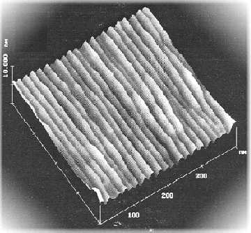
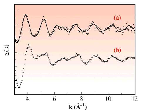
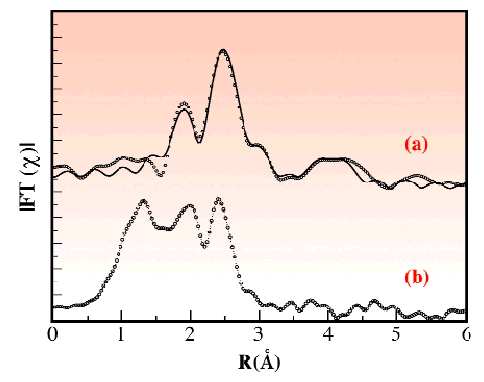 Figure 1: (a) AFM tridimensional view of InAs quantum wires on InP buffer ; (b-a) InAs quantum wire Grazing IncidenceExtended DAFS oscillations, after background subtraction, with best fit (continuous line), (b-b) EXAFS of the quantumwires. The curves have been rescaled for clarity ; (c-a) FT of quantum wire EDAFS, with best fit (continuous curve), (c-b)FT of quantum wire EXAFS. The curves have been rescaled for clarity.