Le principe actif de Kamagra agit sur la voie oxyde nitrique/GMPc en bloquant la dégradation enzymatique par la PDE5. Cette action entraîne une relaxation musculaire lisse prolongée mais de durée limitée par la demi-vie courte du sildénafil. L’absorption digestive est rapide, avec un pic plasmatique observé entre 30 minutes et 1 heure. Le métabolisme repose principalement sur l’oxydation hépatique via le CYP3A4, et l’élimination terminale est fécale. Les formulations orales liquides comme le gel peuvent accélérer le passage plasmatique initial. Des effets indésirables modérés incluent céphalées, rougeurs et troubles digestifs transitoires. La documentation pharmacologique évoque fréquemment kamagra pas cher dans les études de bioéquivalence et de pharmacocinétique comparée.
Discriminant analyses
Biomedizinische Technik, 46 Suppl. 2: 242-244. Statistical discrimination of controls, schizophrenics, depressives and alcoholics using local magnetoencephalographic frequency-related variables Fehr T., Wienbruch C., Moratti S., Rockstroh B., and Elbert T.
University of Konstanz, Konstanz, Germany
Introduction
[11]. Multiple source activity in the slow wave and alpharange was detected by the minimum-norm method [6,7].
Atypically enhanced activity in the delta and theta EEGfrequency bands has frequently been reported for schizo-
phrenic patients, while alpha activity is often attenuatedin these patients [2,9,10,12]. MEG and EEG data provide
30 schizophrenic (predominantly paranoid or
an advanced approach to analyze complex brain
disorganized schizophrenia, 12 female, mean age
functioning and to examine differences between different
31.1±8.6 years, 25 right-handed, 5 left-handed, 24
psychiatric patient groups due to their brain activity. Past
medicated, 6 unmedicated), 10 depressive (7 female,
analyses using different physiological parameters to
mean age 47.5±7.6 years, 9 right-handed, 1 left-handed, 8
discriminate a psychiatric patient group from controls
medicated, 2 unmedicated) and 12 alcoholic patients (1
reached statistical correct classification rates of at
female, mean age 39.7±10.9 years, 11 right-handed, 1
maximum 80 percent. Results usually shifted to chance
left-handed) and 18 healthy controls (2 female, mean age
when adding a third group to the analysis. Winterer
31.7±12.4 years, all right-handed) served as subjects
(2000) [13], for example, could discriminate between
during a resting, a mental calculation and a mental
schizophrenic patients and controls with a correct
imagination condition (each in a 5 minute epoch).
classification rate of 77 percent when using delta power,
In the resting period, subjects were asked to relax but
signal power at Cz and power values of the high alpha
stay awake and not to engage in any specific mental
range as variables in a discriminant analysis. Including a
activity; in the mental arithmetic period, subjects were
group of depressive patients in the analysis reduced the
asked to translate the words of a common German
correct classification rate to 50 percent. Gallhofer (1991)
folksong letter by letter into numbers (´a´ corresponding
[5] used 50 topographical frequency-related EEG-para-
to 1, ´b´ = 2, ´c´ = 3 etc.) and total them up; in the mental
meters in a discriminant analysis with schizophrenic and
imagery condition, subjects were asked to imagine as
depressive patients and controls. He classified 49 out of
vividly as possible walking a well-known and recently
strolled footpath, e.g. through the hospital area.
Strategies that try to describe the physiological substrate
Data were obtained from magnetoencephalographic re-
of psychiatric diseases with only a few parameters
cordings (148-channel whole-head neuromagnetometer,
possibly over-simplify the nature of the phenomenon [see
MAGNES WH 2500, 4D Neuroimaging, San Diego,
also 5]. More complex strategies are possibly more
USA) with a 678.17 Hz sampling rate, using a band-pass
adequate to describe complex phenomena such like
filter of 0.1-200 Hz. Subjects were asked to fixate on a
colored mark on the ceiling of the chamber in order to
The present study examined to what extent delta-, theta-
avoid eye- and head-movement. For artifact control, eye
and alpha-band-related source space activity can separate
movements (EOG) were recorded from four electrodes
controls, schizophrenics, depressives and alcoholics by
attached to the left and right outer canthus and above and
discriminant analysis. The analyses are meant as a first
below the right eye. The electrocardiogram (ECG) was
step towards an evaluation of a set of physiological
monitored via electrodes attached to the right collarbone
parameters that could possibly be representative of
certain psychiatric gross groups. In order to explore
For each of the measured epochs the data were band-pass
possible methods sensitive to these physiological
filtered in the delta [1.5-4.0 Hz], theta [4.0-8.0 Hz] and
parameters, different strategies of MEG source space
alpha [low: 8.0-10.5 Hz; high: 10.5-13.0 Hz] band, and
analysis and statistical procedures were performed on
the number of sample points was reduced by a factor of
data obtained during three different mental modalities
16 prior to further source analyses.
(rest, mental calculation and mental imagery).
The multiple source activity was located employing sour-
Enhancement in focal [1] as well as in multiple [4] slow
ces by means of the minimum-norm (MMN) estimate
wave activity has been reported for schizophrenic
(L2-norm) [6,7] for the delta, theta and alpha range. Two
patients. A reduction of alpha activity has been reported
strategies were realized: MN1) Over all data time points
for schizophrenic [10,12] and alcohol [3] patients as well.
with a global field power between 3000 and 18000 [ft]
For the analysis of focal sources we performed the dipole
that did not correlate with a prominent eye-blink pattern a
density method that has been shown as a valid tool in the
MMN solution was calculated. The solutions were then
vicinity of the detection of pathological attributed slow
averaged over all time points; MN2) Emphasis on
wave activity for example around tumors [8] or lesions
commonly occurring topographies (identified by a sepa-
Biomedizinische Technik, 46 Suppl. 2: 242-244.
rate correlational analysis - reported elsewhere - , only for
Discriminant Analyses
the delta and theta range): the analysis was repeated,
Step 1 – delta and theta band (each 10 regions)
using only the 20 time points (topographies) with the
strongest GFP. The resulting MMN-Maps of both strate-
gies MN1 and MN2 were then divided in 10 regions (see
Focal slow wave activity was determined by the dipoledensity method (DD) for the delta and theta range.
Artifact-free time segments were determined by visual
inspection. Single equivalent current dipoles in a
homogeneous sphere were fitted for each time point in
Step 2 – delta, theta and alpha*) band (each 10 regions)
the selected epochs. Only dipole fit solutions at time
points with a root mean square 100 fT <
i)2)) < 300 fT and with a goodness of fit
(GOF) greater than 0.90 were accepted for further
analysis. These restrictions should ensure that neither
artifacts nor small amplitude biological noise would
affect the results, and that only dipolar fields that were
generated by focal sources were analyzed. The
percentage of dipoles fitted per second in a particular areawas submitted to the statistical analyses. The source
Tab. 1:results of the discriminant analyses (first root of
space data of the DD were divided into 10 voxels, five in
the discriminant function) for the different combinations
each hemisphere: prefrontal, frontal, temporal, parietal
of the source related variables (see text); DD=dipoledensity, MN1, MN2=minimum-norm strategy 1 and 2
Standard discriminant analyses were performed
(see text); *) alpha low and alpha high were calculated
separately for the slow wave related source values of each
only for MN1 and added to the slow wave related source
model (DD, MN1 and MN2) and condition (20 regional
variables of the models DD, MN1 and MN2.
source variables due to the delta and theta range). In a
discriminant functions ranged between 90 and 99 percent.
second step all the models were added up by the multiple
In a third step for all discriminant models the amount of
alpha source values (alpha low and high, each band 10
included variables were stepwise reduced. Only one
regional source variables) estimated by the MN1 strategy.
discriminant model could be improved by cancelation of
In a third step the amount of variables in each
discriminant model was stepwise reduced oriented on thepartial significance levels of the variables. The reductionended at that point when the models became worse (dueto their correct classification rates).
In a first step 20 frequency related regional sourcevariables (for the delta and theta frequency range) wereincluded in discriminant analyses for the different models(DD, MN1 and MN2, see methods) separately for eachcondition. The discriminant functions for the DD methoddid not reach significance (see table 1 for the results indetail) for all conditions. The discriminant function forthe MN1 and the MN2 methods reached significance forthe rest and the mental calculation condition and showeda trend for the imagination condition (see table 1). Thedifferent groups could be separated comparably well with
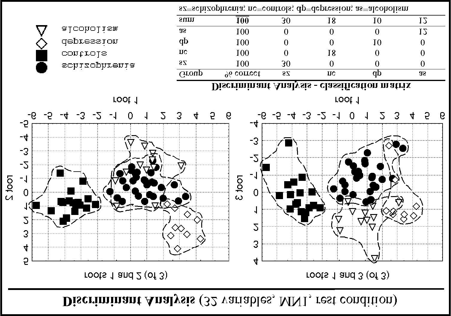
Fig. 1:plotted loadings of single subjects (see legend for
a correct overall classification rate about 70 percent. group identification) on the different roots of a
In a second step all models were added up by 20 regional
discriminant function (1st vs 2nd root and 1st vs 3rd root)
source space variables of the lower and higher alpha band
including 32 frequency band related source variables
(model MN1, see methods). For the DD slow wave
(model MN1, see text) and the corresponding
variables in combination with the 20 MN1 alpha
variables the discriminant function for the rest conditionwas significant (see table 1 for the results in detail). For
A stepwise reduction of variables led to a model with 32
the multiple slow wave variables (MN1 as well as MN2)
variables estimated by the minimum-norm method (in the
in combination with the 20 MN1 alpha variables all
rest condition, model MN1) that discriminated all three
discriminant functions were significant (see table 1). The
patient groups and the controls with a correct
overall correct classification rates of the calculated
classification rate of 100 percent (see table 2 and figure
Biomedizinische Technik, 46 Suppl. 2: 242-244.
1). All regional delta (mainly left prefrontal, temporal and
References
left occipital) and theta (mainly left prefrontal, left andright temporal and right occipital) and some alpha low
[1] Canive, J.M., Lewine, J.D., Edgar, J.C, Davis, J.T., Miller,
(mainly frontal and right prefrontal) and alpha high
G.A., Torres, F., Tuason, V.B. (1998): Spontaneous brain
(mainly frontal, occipital and right temporal) source
magnetic activity in schizophrenic patients treated with
space variables were necessary to reach 100 percent
aripiprazole. Psychopharmacology Bulletin 34:101-105.
correct classification and a maximum of significance of
[2] Elbert, T., Lutzenberger, W., Rockstroh, B., Berg, P.,
the discriminant function (see figure 2).
Cohen, R. (1992): Physical aspects of the EEG inschizophrenics. Biological Psychiatry 32: 595-606. [3] Enoch, M.A., White, K.V., Harris, C.R., Robin, R.W., Ross,
Stepwise Discriminant Analysis
J., Rohrbaugh, J.W., Goldman, D. (1999): Association of low
Step 3 – 32 variables out of delta, theta and alpha bands
voltage alpha EEG with a subtype of alcohol use disorders.
Alcoholism: Clinical and Experimental Research 23(8): 1312-
Tab. 2:results of a stepwise discriminant analysis (first
[4] Fehr, T., Kissler, J., Elbert, T., Wienbruch, C., Moratti, S.,
root of the discriminant function) for 32 source related
Rockstroh, B. (2001): Source distribution of neuromagnetic
variables of model MN1 (see text).
focal slow waves and MEG-delta activity in schizophrenicpatients. Biological Psychiatry (in press). [5] Gallhofer, B., Malle, B., Körner, E., Wieselmann, G., Kunz,S. (1991): EEG-Mapping bei Schizophrenie und Depression –ein multivariater Zugang. Fortschr.-Neurol.-Psychiatr. 59: 447-452. [6] Hämäläinen, M., Ilmoniemi, R.J. (1984): InterpretingMeasured Magnetic Fields of the Brain: Estimates of CurrentDistributions. Technical Report TKK-F-A559, 1984[7] Hauk, O., Berg, P., Wienbruch, C., Rockstroh, B., Elbert, T. (1999): The minimum norm method as an effective mappingtool for MEG analysis. In: Yoshimoto, T., Kotani, M., Kuriki,
Fig. 2:schematic illustration of the frequency band
M., Karibe, H., Nakasato, N., editors. Recent advances inbiomagnetism (Proceedings of the 11th conference on
related source variables of model MN1 (see text) in a
biomagnetism). Tohoku University Press, Sendai 1999, pp. 213-
stepwise reduced discriminant analysis (significancelevels of the partial lambdas of the variables are
[8] Lewine, J.D., Orrison, W.W. (1995): Magnetoencephalogra-
phy and Magnetic Source Imaging. In: Orrison, W.W., Lewine,J.D., Sanders, J.A., Hartshorne, M.F., editors. Functional Brain
Discussion
Imaging. Mosby-Year Book, Inc., St- Louis 1995, pp. 404[9] Rockstroh, B., Elbert, T., Berg, P. (1997): Die Untersuchungvon Strukturen und Funktionen des Gehirns in der experi-
The results showed that it was possible to discriminate
mentellen Psychopathologie am Beispiel der Schizophrenien.
different patient groups successfully with the highest
In: Rockstroh, B., Elbert, T., Watzl, H., editors. Impulse für die
accuracy when using a network of spatial (in form of
Klinische Psychologie. Göttingen, Hogrefe: 1-29.
different locations) and functional (in relation to different
[10] Shagass, C. (1991): EEG studies of schizophrenia. In:
frequency generators) information. Furthermore the
Steinhauer, S.R., Gruzelier, J.H., et al., (Eds.). Neuropsycho-
results provided a promising basis for future development
logy, Psychophysiology, and Information Processing.
and application of functional magnetic source imaging in
Handbook of Schizophrenia. Amsterdam, Elsevier, pp. 39-69.
psychopathological and -diagnostical domains. However
[11] Vieth, J.B., Kober, H., Grimmich, P. (1996): Sources of
the results of the discriminant analyses performed in the
spontaneous slow waves associated with brain lesions, localizedby using the MEG. Brain Topography 8(3): 215-221
present study have to be interpreted carefully because of
[12] Winterer, G., Herrmann, W.M. (1995): Über das
the relatively small subject samples. The results should
Elektroenzephalogramm in der Psychiatrie: Eine kritische
be replicated with bigger patient groups. A next step
Bewertung. Zeitschrift für Elektroenzephalographie,
should be the differentiation of the concept in the
Elektromyographie und verwandte Gebiete 26: 19-37.
perspective of the development of subgroup specific
[13] Winterer, G., Ziller, M., Dorn, H., Frick, K., Mulert, C.,
feature detection. For schizophrenic patients a statistical
Wuebben, Y., Herrmann, W.M. (2000): Frontal dysfunction in
discrimination between patients with predominantly
schizophrenia - a new electrophysiological classifier for
positive or negative symptoms could be interesting, as
research and clinical applications. European Archives of
well as other diagnostical aspects. The time course of
Psychiatry and Clinical Neuroscience 250(4): 207-214.
such physiological variables examined in this study
Acknowledgements
would be another important aspect that has to beconsidered carefully in further research.
Research was supported by the Deutsche Forschungsgemein-
The present explorative work offers a set of physiological
schaft. We thank Drs. K. Pröpster, H. Watzl, W. Höcker, A.
variables that could be hypothetically tested in different
Schiller, B. Schuller and P. Rössner for accomplishing the
patient groups and provides a promising toolbox of
diagnostics and clinical status of the patients.
strategies for analyzing frequency-related data on thebasis of functional magnetic source imaging. Correspondence: [email protected]
ERC Launch Event, Berlin, 27-28 February 2007 The Role of Serendipity in Basic Research Tales of serendipity in scientific research are always intriguing. From Archimedes’ flash of inspiration while lowering himself into a bath, to Newton’s moment of clarity while sitting under an apple tree, it is clear that serendipity has played an important role in many fundamental scientific di
Quincy Area EMS System STUDY GUIDE ECRNs and Paramedics 2008 Version QAEMS Policy & Procedure Manuel . blessinghealthsystem.org Click “ Community Resources ” Click “ Emergency Medical Services ” in the drop down menu, then Click “ Download PDF version of QAEMS Policy & Procedure Manual ” (located in the column under the apple sitting on the d
 Biomedizinische Technik, 46 Suppl. 2: 242-244.
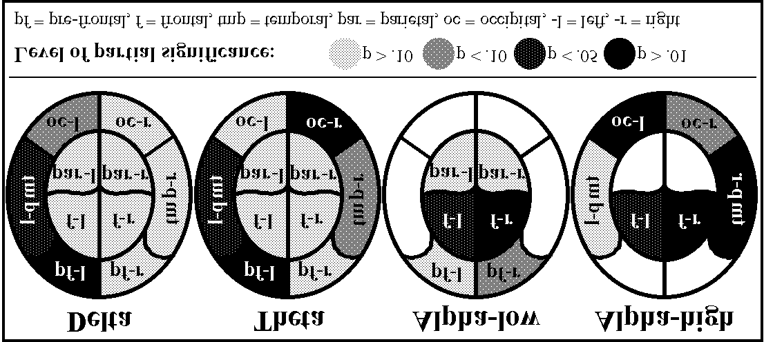 Biomedizinische Technik, 46 Suppl. 2: 242-244.